1. Introduction
Parkinson’s disease (PD) is one of the most common chronic diseases of nervous system in the middle-aged and elderly population (Sale et al.,
2013). Caused by dopaminergic neurons degeneration in nigrostriatal system, PD is clinically characterized by static tremor, muscle rigidity, bradykinesia, and hysteresis, lowering the life quality of PD patients (Tokutake et al.,
2014). Currently, the treatment effects of PD are unsatisfactory and methods are limited to oral administration of
L-dopa, stereotaxic surgeries, and even fetal nigral cell transplantation (Foltynie and Kahan,
2013). However, several clinical trials indicated the inefficacy of the listed therapies evidenced by delayed dyskinesia and insufficient functional recovery in some patients (Zesiewicz et al.,
2007). Therefore, it is necessary and significant to explore alternative therapies.
Glial cell line-derived neurotrophic factor (GDNF) was categorized as a member of a transforming growth factor-β (TGF-β) superfamily (Nielsen et al.,
2009). Previous studies identified GDNF as a strong neuroprotectant because GDNF promoted the development, growth, survival, and maturation of various neurons in the nervous system including the central nervous system (CNS) and the peripheral nervous system (PNS) (Minnich et al.,
2010; Ortiz-Ortiz et al.,
2011). Pre-clinical studies confirmed curative effects of GDNF therapy in the treatment of traumatic brain injury, spinal cord injury, amyotrophic lateral sclerosis, and so on (Grundström et al.,
2000; Bakshi et al.,
2006; Lo et al.,
2008). Moreover, Burke (
2006) found that GDNF promoted survival, differentiation, and repairing of nigrostriatal dopamine neurons. Hebb et al. (
2003) and Hidalgo-Figueroa et al. (
2012) demonstrated that GDNF was potent in attenuating nigrostriatal damage, and thus improved motor symptoms in animal models of PD. In some clinical trials, by intraputaminal infusion, GDNF was administered to PD patients (Patel et al.,
2005). Accumulating results showed improved outcomes without significant side effects (Lang et al.,
2006). Hence, mass production of GDNF is of potential pharmaceutical value in PD treatment.
Astrocyte is the applicable candidate cell secreting GDNF
in vitro mainly through activating signaling transducers including protein kinase Cα (PKCα) (Matsushita et al.,
2008) and cyclic adenosine monophosphate (AMP) responsive element-binding protein (CREB) (Hisaoka et al.,
2008). Compared with the regular 2D culturing system, a 3D culturing system not only realizes a high cell density in a small volume, but also provides an optimistic microenvironment for cells (Li and Cui,
2014). These characteristics enable mass production of the desired factors released from the cultured cells. The physical and chemical properties of the supporting material of the 3D culturing system would impact greatly on the proliferation, differentiation, secretion, and other biological functions of the seeding cells (Zuo and Cai,
2013). In this paper, we developed a novel zirconium oxide (ZrO
2) ceramic foam as the supporting material, which was of high porosity with an open-pore structure, for astrocyte 3D culturing to yield GDNF. The biocompatibility of the material, cell proliferation, and GDNF production were also determined to evaluate its possible application in the pharmaceutical industry.
2. Materials and methods
2.1. ZrO2 ceramic foam preparation
The ZrO
2 foam scaffolds were manufactured and prepared using an ‘impregnation’ method by Drache Umwelttechnik GmbH (Germany). The raw material, ZrO
2 ceramic powder combined with the stabilizer, was mixed with the organic binder agent. The mixture was formed into a ‘slurry’ and absorbed by the organic sponge. Then, the sponge saturated with the slurry was sintered at 1200 °C for about 2 h to decompose sponge skeleton and guarantee the rigidity of the foam ligament.
2.2. Foam physical parameter measurement
Three sets of foam samples with different pore densities (
ω=10, 20, and 30 pores per linear inch (PPI)) were provided by the manufacturer in this study and the nominal foam porosity was 0.90. Some key physical parameters including foam porosity (
ε, volume fraction of void pores), pore size (
d
p), fiber diameter (
d
f), volumetric interfacial area of the ligaments (
a
if), and micro surface area of the material (
a
sf) were determined in this study. It was difficult to measure a typical pore size for a specific pore density since the cells are randomly distributed and most cells seem to be hexagonal, pentagonal, or circular. Hence, we determine pore size and fiber diameter by calculating the arithmetic average value of five different pore sizes and fiber diameters for each foam sample to guarantee the accuracy. The interfacial area of the ligaments (
a
if) is dependent on the pore size (
d
p) and fiber diameter (
d
f). The interfacial area of the foam structure can be calculated by
a
i
f
=
3
π
d
f
/
d
p
2
.
We characterized the micro pore structure by a nitrogen sorption experiment with manometric technique (Autosorb IQ). The sample was degassed by a vacuum molecular pump for 12 h at the atmospheric temperature of 473.15 K before the absorption. Then, the absorption procedure was completed in a liquid nitrogen atmosphere (77 K). The volume and the micro surface area were deduced using the Brunauer-Emmett-Teller (BET) sorption theory based on the measured data.
2.3. Primary astrocyte cell culturing and seeding
Primary astrocytes were harvested from cerebral cortices of neonatal Sprague-Dawley (SD) rats (Animal Experimental Center, Xi’an Jiaotong University, China) according to the protocol described by Cho et al. (
2010). The animal experimental procedures were approved and authorized (License No. 2014-O59) by the Ethics Committee of Experimental Animals in Xi’an Jiaotong University, China. After digestion, dissociated cortical cells were suspended in Dulbecco’s modified Eagle medium (DMEM; Gibco) containing 10% fetal bovine serum (FBS; Gibco), 2 mmol/L
L-glutamine (Invitrogen), and 100 U/ml penicillin-streptomycin (Invitrogen). Cells were cultured in 25-cm
2 culture flasks (Corning). After agitation and culture medium replacement, non-astrocytic cells (neurons and microglia) were detached and removed from the culturing system. After 12–14 d cell culture, a monolayer of purified (more than 95% purity, testified by glial fibrillary acidic protein (GFAP) staining) primary astrocytes could be acquired.
The ceramic foam scaffolds were washed by aviation kerosene and then by ethanol absolute, sterilized prior to cell seeding. The interior wall of each well in a six-well plate (Corning) was coated with Sigmacote (Sigma-Aldrich) to eliminate cell attachment to the wall. A laser cutter was used to modify the shape of the ceramic foam (cylinder with diameter of 3.5 cm and height of 1.5 cm) to fit in the culturing wells of the six-well plate. As the diameters of the foam cylinder and the well were the same, the foam was fixed to the well by contracting the interior wall of the wells. The same amount of cells (6×10
6 cells/20 ml) was seeded to the foam scaffold with different pore densities (10, 20, and 30 PPI) soaked in the culturing medium and to bare medium as a control in six-well plates. The culturing medium in the wells of the culturing plate was replaced every two days. The astrocytes cultured in the six-well plates were harvested by digestion. The same amount of harvested cells from each group was used for the subsequent experiments including cell viability assay, cell proliferation assay, enzyme-linked immunosorbent assay (ELISA), real-time polymerase chain reaction (PCR), and Western blotting.
2.4. Scanning electron microscopy observation
Samples of ceramic foams or foams with cells embedded were collected and prepared for scanning electron microscope (SEM) observation. Samples were fixed in glutaraldehyde (2.5%) for 2 h, then washed by distilled water, and then immersed in OsO
4 (4%) for 1 h. After dehydration by a graded ethanol solution (25%, 50%, 75%, 80%, 90%, and 100%), the samples were dried and coated with gold-palladium by a JFC-1600 auto sputter coater (JEOL, Japan), and then observed with a TM-1000 SEM (Hitachi, Japan).
2.5. Cell viability assay
The biocompatibility of the ceramic foam was evaluated by MTT (3-[4,5-dimethylthiazol-2-yl]-2,5 diphenyltetrazolium bromide) assay after culturing. MTT reagent (50 μl) was added to each well and incubated in 10% CO
2 at 37 °C for 4 h. The formed crystals were dissolved in solution (1% Triton-X 100 and 0.01 mol/L HCl in isopropanol). The absorbance at 570 nm of each well was read by a plate reader (Bio-Rad). The viable cell percentage was expressed as a percentage of viable cells in each culturing system.
2.6. Cell proliferation examination
The proliferation of astrocytes was evaluated by DNA content assessment. DNA was extracted from all cells collected from each culturing system by using a mammalian genomic DNA extraction kit (Beyotime Biotechnology, China). The content of DNA was then determined using a Quant-iT™ PicoGreen double-stranded DNA (dsDNA) assay kit (Invitrogen). All procedures above followed the instructions of the manufacturers.
2.7. ELISA assessment of GDNF production
The supernatants from the astrocytes culturing system were collected for ELISA assay using a rat GDNF ELISA kit (Promega) according to the protocol provided by the manufacturer. After the absorbance was detected at 450 nm using a plate reader (Bio-Rad), the concentrations of GDNF in the supernatants were then calculated.
2.8. Real-time quantitative PCR
Total RNA was extracted from the cultured astrocytes with the RNAfast 200 kit (Fastagen), and then reverse-transcribed to complementary DNA (cDNA) with the PrimeScript RT reagent kit (TaKaRa, Japan) according to the protocols provided by the manufacturers. Real-time quantitative PCR was performed using the SYBR Premix Ex Taq™ II (TaKaRa, Japan) and Prism 7500 real-time detection system (Applied Bio-Systems). Oligonucleotide primers for GDNF and β-actin are listed in Table
1. Relative mRNA expression levels (normalized to β-actin) were calculated by relative cycle threshold method using Bio-Rad IQ5 software (Ver 1.0, Bio-Rad).
Table 1
Primers for real-time PCR
| Barley variety |
Primer sequence (5'–3') |
Size (bp) |
| GDNF |
F: GACTCCAATGTGCCCGAAGA |
394 |
| R: CCTACCTTGTCACTTGCTAGCC |
| β-actin |
F: TGGCACCCAGCACAATGAA |
186 |
| R: CTAAGTCATAGTCCGCCTAGAAGCA |
2.9. Western blotting
The same amount of cultured astrocytes (6×10
6) was lysed and homogenized in an RIPA (radio-immunoprecipitation assay) lysis buffer system with phenylmethylsulfonyl fluoride (PMSF) (Santa Cruz). The resultant supernatants were separated by centrifugation at 12 500
g at 4 °C for 10 min. The protein concentration in each sample was detected by the BCA protein assay kit (Santa Cruz). After subjecting to a 1× sodium dodecyl sulfate polyacrylamide gel electrophoresis (SDS-PAGE) loading buffer, sample protein was then separated by vertical electrophoresis in an SDS-polycrylamide gel (10%). Separated protein was then transferred to a polyvinylidene fluoride (PVDF) membrane. The blots were probed by specific antibodies against the proliferating cell nuclear antigens (PCNA; 5000×; Abcam), PKCα (5000×; Abcam), total CREB (5000×; Cell Signaling Tech.), phospho CREB (5000×; Cell Signaling Tech.), GDNF (5000×; Abcam), and β-actin (2000×; Santa Cruz) at 4 °C for 10 h. After being washed by Tris-buffered saline (TBS)-Tween 20 (0.02%), a second antibody conjugated to horseradish peroxidase (HRP; Santa Cruz) was employed to incubate the membrane. The membranes were developed with Super Signal West Pico chemiluminsecence reagent (Thermo Scientific) and visualized on X-ray films.
2.10. PKCα translocation evaluation
The same amount of cells (6×10
6) from each culturing system was centrifuged, collected, and lysed. Harvested lysates were centrifuged at 500
g for 10 min, and then the supernatant was further centrifuged at 100 000
g at 4 °C for 1 h. The resultant supernatant was collected as a cytosolic fraction. The resultant pellet was resuspended in a protein extraction buffer, and then centrifuged at 100 000
g for 1 h. The resultant supernatant was collected as a membrane fraction. The translocation of PKCα was evaluated by the expression level of PKCα in the membrane fraction detected by Western blotting.
3. Results
3.1. Physical parameters of ZrO2 ceramic foam
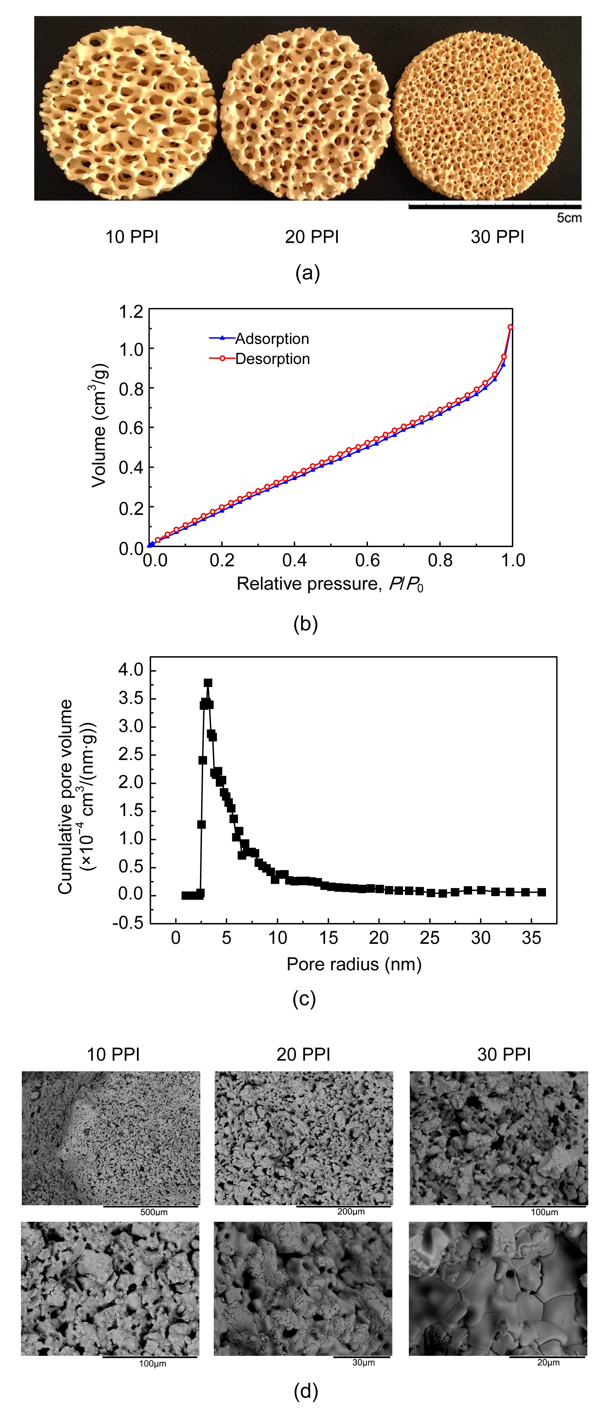
The macrostructures and microstructures of the typical ZrO
2 foams used in this study are shown in Figs.
1a and 1d. It can be observed that the foam consists of cell-like pores which are formed by inner-connected fiber ligaments. The measured and calculated results of porosity, pore size, fiber diameter, and interfacial area are listed in Table
2. The micro-volume increased in linear relation with the relative pressure from 0 to 0.93 (Fig.
1b). This indicates a relative uniform pore distribution and sustainable pore characteristic for absorption (desorption). The volume encounters a dramatic increase with the pressure ratio of 0.93–1.00. Fig.
1c shows the variation of cumulative pore volume distribution with the cell width, which shows the pore diameter ranging 2.5–10.0 nm with the maximum pore distribution at the peak size of 3.169 nm.
Fig.1
Physical properties of ZrO2 ceramic foam
(a) Macrostructures of ZrO2 ceramic foams with three different pore densities (10, 20, and 30 PPI) captured by an optical camera; (b) Nitrogen absorption results of the micro-volume as the function of the pressure ratio for the ZrO2 foam; (c) Variation of cumulative pore volume distribution with the cell width by using nitrogen sorption method; (d) Different magnification of fiber surfaces of ZrO2 foam captured by scanning electron microscopy
Table 2
Physical parameters of ZrO2 ceramic foam
| Pore density (PPI) |
Pore size (mm2) |
Fiber diameter (mm) |
Interfacial area (m−1) |
| 10 |
2.94 |
0.357 |
0.389 |
| 20 |
1.73 |
0.029 |
0.659 |
| 30 |
0.78 |
0.094 |
0.456 |
3.2. ZrO2 ceramic foam astrocyte culturing system elevated GDNF yield
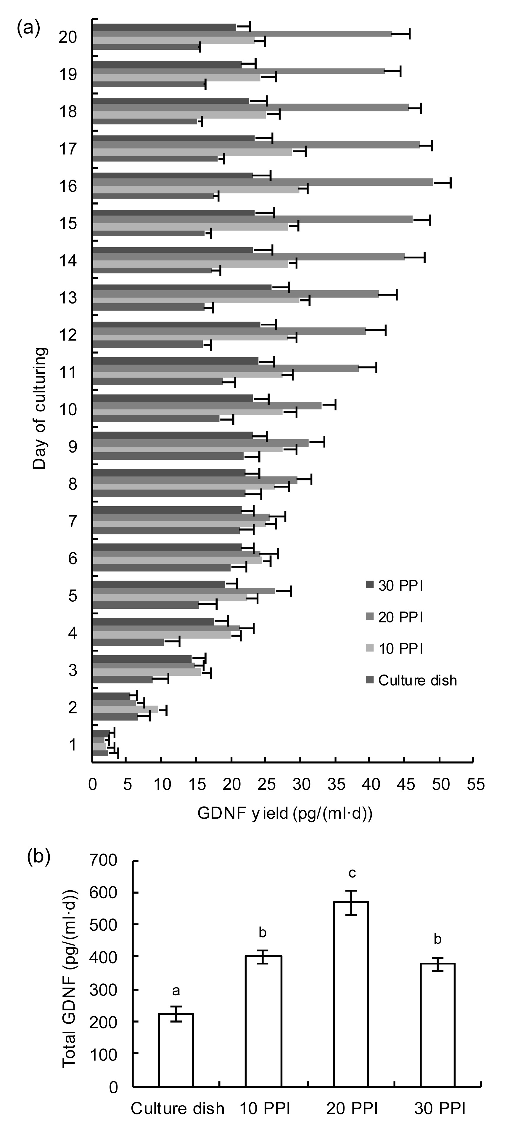
Fig.
2 shows the yields of GDNF from each astrocytes culturing system over 20 d starting from seeding. The productivity of GDNF increased significantly and was sustained through the 20-d period in the ceramic foam culturing system compared with regular 2D culturing system whose GDNF productivity began to fall after 10-d culturing. In the ceramic foam culturing system, foam with pore density of 20 PPI exhibited the highest average GDNF yield during the 20-d observation compared with that of 10 PPI and 30 PPI.
Fig.2
GDNF productivity in ZrO2 ceramic foam astrocyte culturing system
Daily GDNF yield (a) and total production of GDNF (b) from different culturing systems including regular culture dish and ZrO2 ceramic foam 3D culturing systems with different pore densities (10, 20, and 30 PPI) during the 20-d culturing. Values are presented as mean±SD with different letters above the bars showing P<0.005
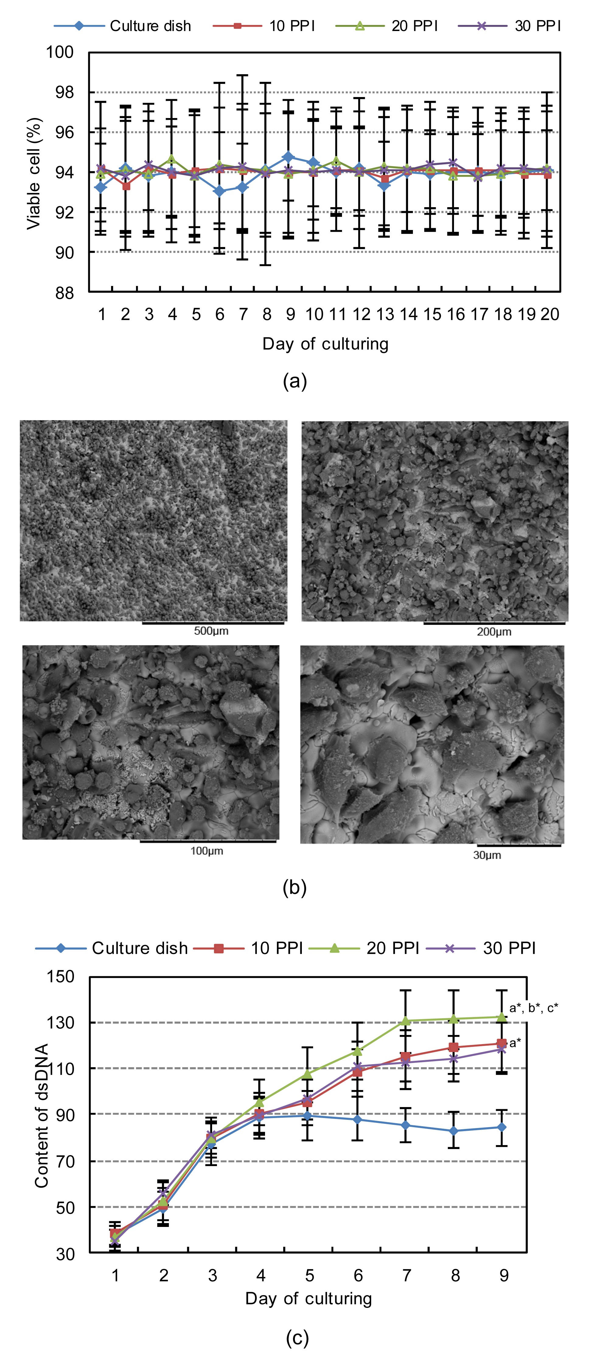
3.3. ZrO2 ceramic foam was biocompatible and enhanced astrocytes proliferation in limited volume
There were no significant differences of the viable cell percentage in each culturing system evidenced by MTT assay (Fig.
3a). The attachment of seeded astrocytes to ceramic foam is observed by SEM (Fig.
3b). The dsDNA content was more dramatically elevated in the ceramic foam culturing system than in the regular one (Fig.
3c). dsDNA content increased more significantly in foam with pore density of 20 PPI than of 10 PPI and 30 PPI. Additionally, the elevated expression of PCNA on Day 8 in astrocytes in the ceramic foam culturing system is shown in Fig.
4.
Fig.3
Astrocytes seeded in ZrO2 ceramic foam
(a) Viable cell percentages of seeded astrocytes in different culturing systems during 20-d culturing by MTT assay; (b) SEM pictures of astrocytes seeded in ZrO2 ceramic foam; (c) Content of dsDNA extracted from astrocytes seeded in different culturing systems during 20-d culturing. a, b, and c shows values significantly different from culture dish, 10 PPI, and 30 PPI, respectively. *
P<0.005
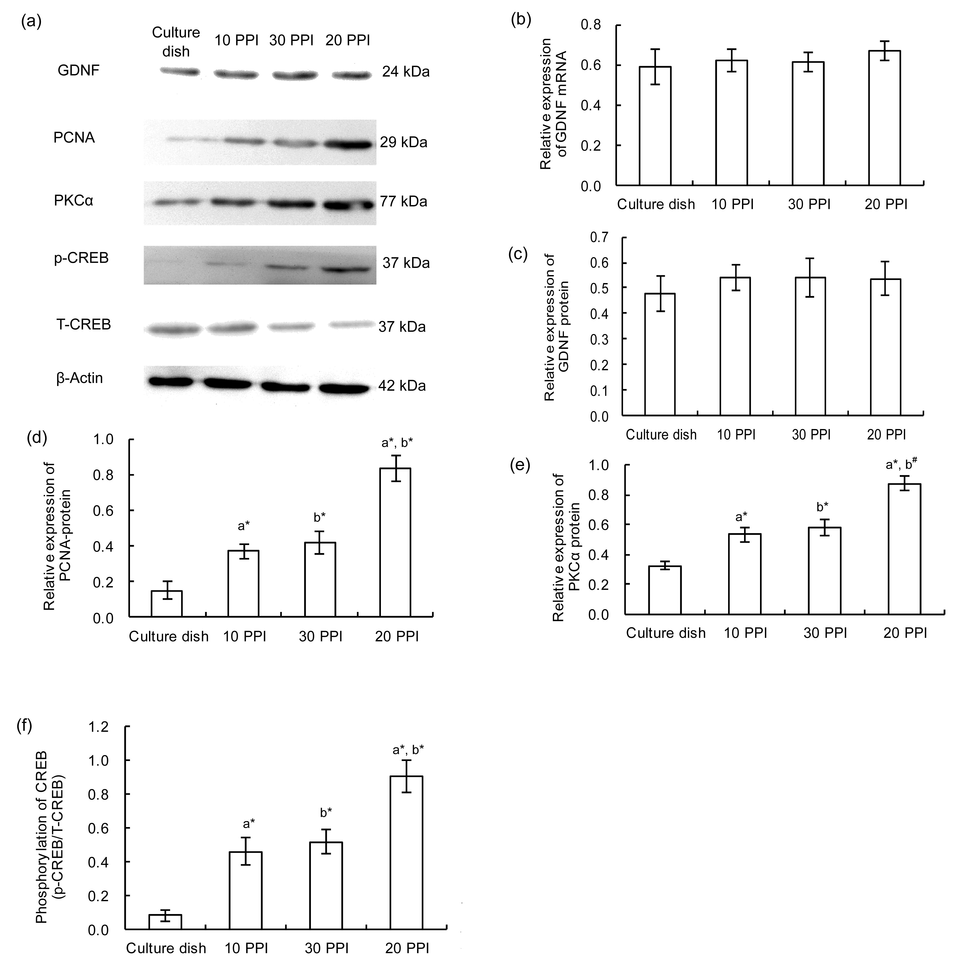
Fig.4
Changes of expressions of molecules in seeded astrocytes on Day 8 of culturing
(a) Western blotting of GDNF, PCNA, PKCα, p-CREB, T-CREB, and in astrocytes on Day 8 of culturing in different culturing systems: regular culture dish and ZrO2 ceramic foam 3D culturing systems with pore densities of 10, 20, and 30 PPI. Relative expressions of GDNF mRNA (b), GDNF protein (c), PCNA protein (d), PKCα protein (e), and p-CREB/T-CREB (f) in astrocytes on Day 8 of culturing in each culturing system. Values are presented as mean±SD. a and b above the bars shows values significantly different from culture dish and 10 PPI, respectively. #
P<0.01; *
P<0.005
3.4. ZrO2 ceramic foam cell culturing pattern stimulated synthesis and secretion of GDNF in astrocytes
On Day 8 during culturing, at both transcriptional and translational levels, there were no significant differences of GDNF expression in the same amount of astrocytes from the ceramic foam culturing system than from the regular one (Fig.
4). Notably, in the system supported by foam, in the same amount of astrocytes from each culturing system, expression of GDNF, translocation of PKCα, and phosphorylation of CREB increased significantly in the ceramic foam culturing system compared with the regular one; this was more dramatic in the system supported by foam with pore density of 20 PPI than of 10 PPI and 30 PPI.
4. Discussion
The normal structure and function of organs
in vivo are maintained by natural cell alignments, metabolism, complicated cellular interactions, and so on, which rely on the 3D structural nature of biological tissues. Biological active factors, such as cytokines and tumor antibodies, are employed in clinical treatments (Schenck et al.,
2007; Elliot et al.,
2008; Beohar et al.,
2010; von Frenckell and Malaise,
2012). Most of these biological factors could not be acquired by artificial synthesis but through selection and purification from the products generated by specific living cells (Tanaka et al.,
2008). Although these biological products could be harvested from regular 2D cell culturing system, the productivity is not optimistic because of the inadequacy in mimicking the
in vivo culturing environment and the vulnerability to external stimuli of the 2D culturing system (Heckmann et al.,
2008). Hence, we introduced ZrO
2 ceramic foam as a scaffold to support the 3D cell culturing system. Primary astrocytes were cultured in this system, and the production of its final biological products, GDNF, was found to be elevated dramatically compared with the regular 2D culturing system. Mechanically, in this study, we also demonstrated that the elevation of GDNF yield was due to the favorable biocompatibility of ZrO
2 ceramic foam as well as the increased proliferation and stimulated GDNF synthesis in astrocytes seeded in the 3D culturing system.
To achieve a high efficiency, it is desirable that a generating device for biological products produces the highest possible number of cells within a limited volume. In this study, we prepared ZrO
2 ceramic foams with serial pore densities of 10, 20, and 30 PPI. The interfacial specific surface areas of the foams were significantly increased by the foam fiber surfaces within the same volume, indicating that the foams could provide a higher surface area per unit volume. Scanning electronic microscopy observation of the foam showed the features of uneven topography and rough fiber surface, which would facilitate anchoring, adaptation, and attachment of astrocytes to this scaffold. Additionally, the cell viability assay confirmed the favorable response of primary astrocytes after being seeded to the ceramic foams. These featured characteristics of ZrO
2 ceramic foam enabled it to sustain a large cell population to produce more biological products. In this study, we found that the ceramic foam with pore density of 20 PPI was more suitable for astrocytes culturing, by improving the amount of cells. Pore density is one of the determining physical parameters of ceramic foam which would affect the biological properties of cells when ceramic foams are utilized in a cell culturing system. At a given dimension, the foam with higher pore density has more available surface internal area. As the internal surface of the foam is for cell attachment, providing more space for cell growth, more cells would settle in the foam with a higher pore density. As a result, the cell proliferation is boosted. However, as the pore density increases, the flowability of the culturing medium in the ceramic foam is decreased. Thus the oxygen and nutrients in the medium could not penetrate to the deeper space in the foam. Additionally, the internal structure of the foam with high pore density is more complex which would offer obstructions or barriers for cell migration to the deeper space of the foam. As a result, a ‘bare area’ without cells or with few cells could exist in the foam at high pore density.
In this study, we chose astrocytes as seeding cells in the 3D culturing system to produce GDNF. As a member of TGF-β, GDNF was known for its neuroprotective and restorative effects on dopaminergic neurons (Sun et al.,
2005; Wu et al.,
2008). GDNF acts as a ligand to induce the activation of several transmembrane receptors in dopaminergic neurons such as the ligand binding component GDNF-family receptor α-1 (GFRα1). Then the down-stream signaling pathways such as mitogen-activated protein kinase pathway are activated and this determines the survival of the neurons. GDNF treatment was considered a feasible and promising treatment strategy in curing PD (Foltynie and Kahan,
2013). In an animal model of PD, both GDNF ventricular infusion and GDNF secreting cell transplantation showed positive therapeutic effects, which was evidenced by an improved behavioral score and rehabilitated dopaminergic neurons in 6-hydroxydopamine lesioned striatum (Shingo et al.,
2002; Yasuhara et al.,
2005). The achievements for GDNF clinical treatment are also encouraging since the first successful intraputaminal GDNF infusion treatments in PD patients in 2003 (Gill et al.,
2003). It was reported that direct administration of GDNF into putamen in PD patients improved their unified Parkinson’s disease rating scale (UPDRS) scores in the following year off medication without any serious side effects (Yasuhara et al.,
2007). Remarkable functional recovery in PD patients was found in accordance with pathological changes of dopaminergic fibers sprouting in PD patients receiving GDNF infusion treatment (Gill et al.,
2003; Patel et al.,
2005). As the technique of cell transplantation is relatively complex and its feasibility is currently in doubt, the utility of direct intraputaminal/intraventricular GDNF injection is the appropriate alternative strategy in curing PD. Future pharmaceutical demands of GDNF can be predicted. A device or system, such as the 3D astrocytes culturing system in this study, for massive production of GDNF would be of potential practical value.
The yield of GDNF in the 3D astrocyte culturing system in this study was considerable. Evidenced by increased dsDNA level and PCNA expression, the proliferation of primary astrocytes in the ZrO
2 ceramic foam supported 3D culturing system was dramatically stimulated. Assuming that each single astrocyte secretes and releases an equal amount of GDNF, a larger viable cell population per unit volume should be responsible for a substantial GDNF production from the system. The optimal pore density for astroycyte proliferation was 20 PPI rather than 30 PPI or 10 PPI. The activation of several cellular signaling pathways is also responsible for GDNF production. In several previous studies, stimulated GDNF release from astrocytes was PKCα-dependent (Matsushita et al.,
2008). CREB was reported as another important activator in promoting GDNF production in cultured astrocytes (Parsadanian et al.,
2006; Hisaoka et al.,
2008; Golan et al.,
2011). Generally, an increased PKCα translocation level and CREB phosphorylation level indicate the activation of PKCα and CREB related pathways. In the present study, notably, on Day 8 during culturing, when under the similar conditions of cell amount and GDNF productivity, significantly increased PKCα translocation and CREB phosphorylation were found in astrocytes from the 3D culturing system compared with the regular 2D one. This result indicated that the 3D culturing pattern stimulated GDNF generating signaling pathways in cultured astrocytes.
5. Conclusions
Our findings suggested that: (1) the ZrO
2 ceramic foam was biocompatible when supporting primary astrocyte 3D culturing; (2) the yield of GDNF of ZrO
2 ceramic foam based 3D astrocytes culturing system overwhelmed that from the regular 2D culturing system; (3) the considerable productivity of GDNF in the 3D system was due to elevation of astrocyte proliferation and the stimulation of GDNF secretion pathways from astrocytes.
* Project supported by the National Natural Science Foundation of China (Nos. 81171262 and 81371473)Compliance with ethics guidelines Zhong-wei LIU, Wen-qiang LI, Jun-kui WANG, Xian-cang MA, Chen LIANG, Peng LIU, Zheng CHU, and Yong-hui DANG declare that they have no conflict of interest.References
[1] Bakshi, A., Shimizu, S., Keck, C.A., 2006. Neural progenitor cells engineered to secrete GDNF show enhanced survival, neuronal differentiation and improve cognitive function following traumatic brain injury.
Eur J Neurosci, 23(8):2119-2134.


[2] Beohar, N., Rapp, J., Pandya, S., 2010. Rebuilding the damaged heart: the potential of cytokines and growth factors in the treatment of ischemic heart disease.
J Am Coll Cardiol, 56(16):1287-1297.
[3] Burke, R.E., 2006. GDNF as a candidate striatal target-derived neurotrophic factor for the development of substantia nigra dopamine neurons.
Parkinsons Disease and Related Disorders, Springer-Verlag/Wien,:41-45.
[4] Cho, K.S., Park, S.H., Joo, S.H., 2010. The effects of IL-32 on the inflammatory activation of cultured rat primary astrocytes.
Biochem Biophys Res Commun, 402(1):48-53.
[5] Elliot, M.J., Maini, R.N., Feldmann, M., 2008. Treatment of rheumatoid arthritis with chimeric monoclonal antibodies to tumor necrosis factor α.
Arthritis Rheum, 58(S2):S92-S101.
[6] Foltynie, T., Kahan, J., 2013. Parkinson’s disease: an update on pathogenesis and treatment.
J Neurol, 260(5):1433-1440.
[7] Gill, S.S., Patel, N.K., Hotton, G.R., 2003. Direct brain infusion of glial cell line-derived neurotrophic factor in Parkinson disease.
Nat Med, 9(5):589-595.
[8] Golan, M., Schreiber, G., Avissar, S., 2011. Antidepressants elevate GDNF expression and release from C
6 glioma cells in a β-arrestin1-dependent, CREB interactive pathway.
Int J Neuropsychopharmacol, 14(10):1289-1300.
[9] Grundstrm, E., Lindholm, D., Johansson, A., 2000. GDNF but not BDNF is increased in cerebrospinal fluid in amyotrophic lateral sclerosis.
NeuroReport, 11(8):1781-1783.
[10] Hebb, A.O., Hebb, K., Ramachandran, A.C., 2003. Glial cell line-derived neurotrophic factor-supplemented hibernation of fetal ventral mesencephalic neurons for transplantation in Parkinson disease: long-term storage.
J Neurosurg, 98(5):1078-1083.
[11] Heckmann, L., Fiedler, J., Mattes, T., 2008. Interactive effects of growth factors and three-dimensional scaffolds on multipotent mesenchymal stromal cells.
Biotechnol Appl Biochem, 49(3):185-194.
[12] Hidalgo-Figueroa, M., Bonilla, S., Gutierrez, F., 2012. GDNF is predominantly expressed in the PV+ neostriatal interneuronal ensemble in normal mouse and after injury of the nigrostriatal pathway.
J Neurosci, 32(3):864-872.
[13] Hisaoka, K., Maeda, N., Tsuchioka, M., 2008. Antidepressants induce acute CREB phosphorylation and CRE-mediated gene expression in glial cells: a possible contribution to GDNF production.
Brain Res, 1196:53-58.
[14] Lang, A.E., Gill, S., Patel, N.K., 2006. Randomized controlled trial of intraputamenal glial cell line-derived neurotrophic factor infusion in Parkinson disease.
Ann Neurol, 59(3):459-466.
[15] Li, Z., Cui, Z., 2014. Three-dimensional perfused cell culture.
Biotechnol Adv, 32(2):243-254.
[16] Lo, W.C., Hsu, C.H., Wu, A.T., 2008. A novel cell-based therapy for contusion spinal cord injury using GDNF-delivering NIH
3T
3 cells with dual reporter genes monitored by molecular imaging.
J Nucl Med, 49(9):1512-1519.
[17] Matsushita, Y., Nakajima, K., Tohyama, Y., 2008. Activation of microglia by endotoxin suppresses the secretion of glial cell line-derived neurotrophic factor (GDNF) through the action of protein kinase c α (PKCα) and mitogen-activated protein kinases (MAPKs).
J Neurosci Res, 86(9):1959-1971.
[18] Minnich, J.E., Mann, S.L., Stock, M., 2010. Glial cell line-derived neurotrophic factor (GDNF) gene delivery protects cortical neurons from dying following a traumatic brain injury.
Restor Neurol Neurosci, 28(3):293-309.
[19] Nielsen, J., Gotfryd, K., Li, S., 2009. Role of glial cell line-derived neurotrophic factor (GDNF)-neural cell adhesion molecule (NCAM) interactions in induction of neurite outgrowth and identification of a binding site for ncam in the heel region of GDNF.
J Neurosci, 29(36):11360-11376.
[20] Ortiz-Ortiz, M.A., Moran, J.M., Ruiz-Mesa, L.M., 2011. Protective effect of the glial cell line-derived neurotrophic factor (GDNF) on human mesencephalic neuron-derived cells against neurotoxicity induced by paraquat.
Environ Toxicol Pharmacol, 31(1):129-136.
[21] Parsadanian, A., Pan, Y., Li, W., 2006. Astrocyte-derived transgene GDNF promotes complete and long-term survival of adult facial motoneurons following avulsion and differentially regulates the expression of transcription factors of AP-1 and ATF/CREB families.
Exp Neurol, 200(1):26-37.
[22] Patel, N.K., Bunnage, M., Plaha, P., 2005. Intraputamenal infusion of glial cell line-derived neurotrophic factor in PD: a two-year outcome study.
Ann Neurol, 57(2):298-302.
[23] Sale, P., de Pandis, M.F., Vimercati, S.L., 2013. The relation between Parkinson’s disease and ageing. Comparison of the gait patterns of young Parkinson’s disease subjects with healthy elderly subjects.
Eur J Phys Rehabil Med, 49(2):161-167.
[24] Schenck, M., Borgermann, C., vom Dorp, F., 2007. Proapoptotic antibodies as new anticancer drugs.
Der Urologe, (in German),46(9):1262-1265.
[25] Shingo, T., Date, I., Yoshida, H., 2002. Neuroprotective and restorative effects of intrastriatal grafting of encapsulated GDNF-producing cells in a rat model of Parkinson’s disease.
J Neurosci Res, 69(6):946-954.
[26] Sun, M., Kong, L., Wang, X., 2005. Comparison of the capability of GDNF, BDNF, or both, to protect nigrostriatal neurons in a rat model of Parkinson’s disease.
Brain Res, 1052(2):119-129.
[27] Tanaka, Y., Ogasawara, T., Asawa, Y., 2008. Growth factor contents of autologous human sera prepared by different production methods and their biological effects on chondrocytes.
Cell Biol Int, 32(5):505-514.
[28] Tokutake, T., Ishikawa, A., Yoshimura, N., 2014. Clinical and neuroimaging features of patient with early-onset Parkinson’s disease with dementia carrying SNCA p.G51d mutation.
Parkinsonism Relat Disord, 20(2):262-264.
[29] von Frenckell, C., Malaise, M.G., 2012. Targeted therapy in inflammatory disease: cytokines.
Rev Med Liege, 67:22-28.
[30] Wu, X., Chen, P.S., Dallas, S., 2008. Histone deacetylase inhibitors up-regulate astrocyte GDNF and BDNF gene transcription and protect dopaminergic neurons.
Int J Neuropsychopharmacol, 11(8):1123-1134.
[31] Yasuhara, T., Shingo, T., Muraoka, K., 2005. Early transplantation of an encapsulated glial cell line-derived neurotrophic factor-producing cell demonstrating strong neuroprotective effects in a rat model of Parkinson disease.
J Neurosurg, 102(1):80-89.
[32] Yasuhara, T., Shingo, T., Date, I., 2007. Glial cell line-derived neurotrophic factor (GDNF) therapy for Parkinson’s disease.
Acta Med Okayama, 61(2):51-56.
[33] Zesiewicz, T.A., Sullivan, K.L., Hauser, R.A., 2007. Levodopa-induced dyskinesia in Parkinson’s disease: epidemiology, etiology, and treatment.
Curr Neurol Neurosci Rep, 7(4):302-310.
[34] Zuo, D.Q., Cai, Z.D., 2013. Progress in three-dimensional cell culture techniques and its application in bone tumor research.
Chin J Oncol, (in Chinese),35(9):641-644.
 ORCID:
Open peer comments: Debate/Discuss/Question/Opinion
<1>