Full Text:   <3061>

Summary:  <2195>

Suppl. Mater.: 

CLC number: Q78

On-line Access: 2014-05-05

Received: 2013-12-31

Revision Accepted: 2014-04-04

Crosschecked: 2014-04-23

Cited: 6

Clicked: 5754

Citations:  Bibtex RefMan EndNote GB/T7714

-   Go to

Article info.
1.  Introduction
2.  Materials and methods
3.  Results
4.  Discussion
5.  Conclusions
6. Reference List
Open peer comments

Journal of Zhejiang University SCIENCE B 2014 Vol.15 No.5 P.466-473

http://doi.org/10.1631/jzus.B1400001


Early lethality of shRNA-transgenic pigs due to saturation of microRNA pathways* #


Author(s):  Zhen Dai1, Rong Wu1, Yi-cheng Zhao1,2, Kan-kan Wang1, Yong-ye Huang1, Xin Yang1, Zi-cong Xie1, Chang-chun Tu3, Hong-sheng Ouyang1, Tie-dong Wang1, Da-xin Pang1

Affiliation(s):  1. Jilin Provincial Key Laboratory of Animal Embryo Engineering, College of Animal Sciences, Jilin University, Changchun 130062, China; more

Corresponding email(s):   tdongw@sohu.com

Key Words:  MicroRNA pathway, shRNA-transgenic pigs, Classical swine fever virus (CSFV), Blastocyst formation, Early lethality


Zhen Dai, Rong Wu, Yi-cheng Zhao, Kan-kan Wang, Yong-ye Huang, Xin Yang, Zi-cong Xie, Chang-chun Tu, Hong-sheng Ouyang, Tie-dong Wang, Da-xin Pang. Early lethality of shRNA-transgenic pigs due to saturation of microRNA pathways[J]. Journal of Zhejiang University Science B, 2014, 15(5): 466-473.

@article{title="Early lethality of shRNA-transgenic pigs due to saturation of microRNA pathways",
author="Zhen Dai, Rong Wu, Yi-cheng Zhao, Kan-kan Wang, Yong-ye Huang, Xin Yang, Zi-cong Xie, Chang-chun Tu, Hong-sheng Ouyang, Tie-dong Wang, Da-xin Pang",
journal="Journal of Zhejiang University Science B",
volume="15",
number="5",
pages="466-473",
year="2014",
publisher="Zhejiang University Press & Springer",
doi="10.1631/jzus.B1400001"
}

%0 Journal Article
%T Early lethality of shRNA-transgenic pigs due to saturation of microRNA pathways
%A Zhen Dai
%A Rong Wu
%A Yi-cheng Zhao
%A Kan-kan Wang
%A Yong-ye Huang
%A Xin Yang
%A Zi-cong Xie
%A Chang-chun Tu
%A Hong-sheng Ouyang
%A Tie-dong Wang
%A Da-xin Pang
%J Journal of Zhejiang University SCIENCE B
%V 15
%N 5
%P 466-473
%@ 1673-1581
%D 2014
%I Zhejiang University Press & Springer
%DOI 10.1631/jzus.B1400001

TY - JOUR
T1 - Early lethality of shRNA-transgenic pigs due to saturation of microRNA pathways
A1 - Zhen Dai
A1 - Rong Wu
A1 - Yi-cheng Zhao
A1 - Kan-kan Wang
A1 - Yong-ye Huang
A1 - Xin Yang
A1 - Zi-cong Xie
A1 - Chang-chun Tu
A1 - Hong-sheng Ouyang
A1 - Tie-dong Wang
A1 - Da-xin Pang
J0 - Journal of Zhejiang University Science B
VL - 15
IS - 5
SP - 466
EP - 473
%@ 1673-1581
Y1 - 2014
PB - Zhejiang University Press & Springer
ER -
DOI - 10.1631/jzus.B1400001


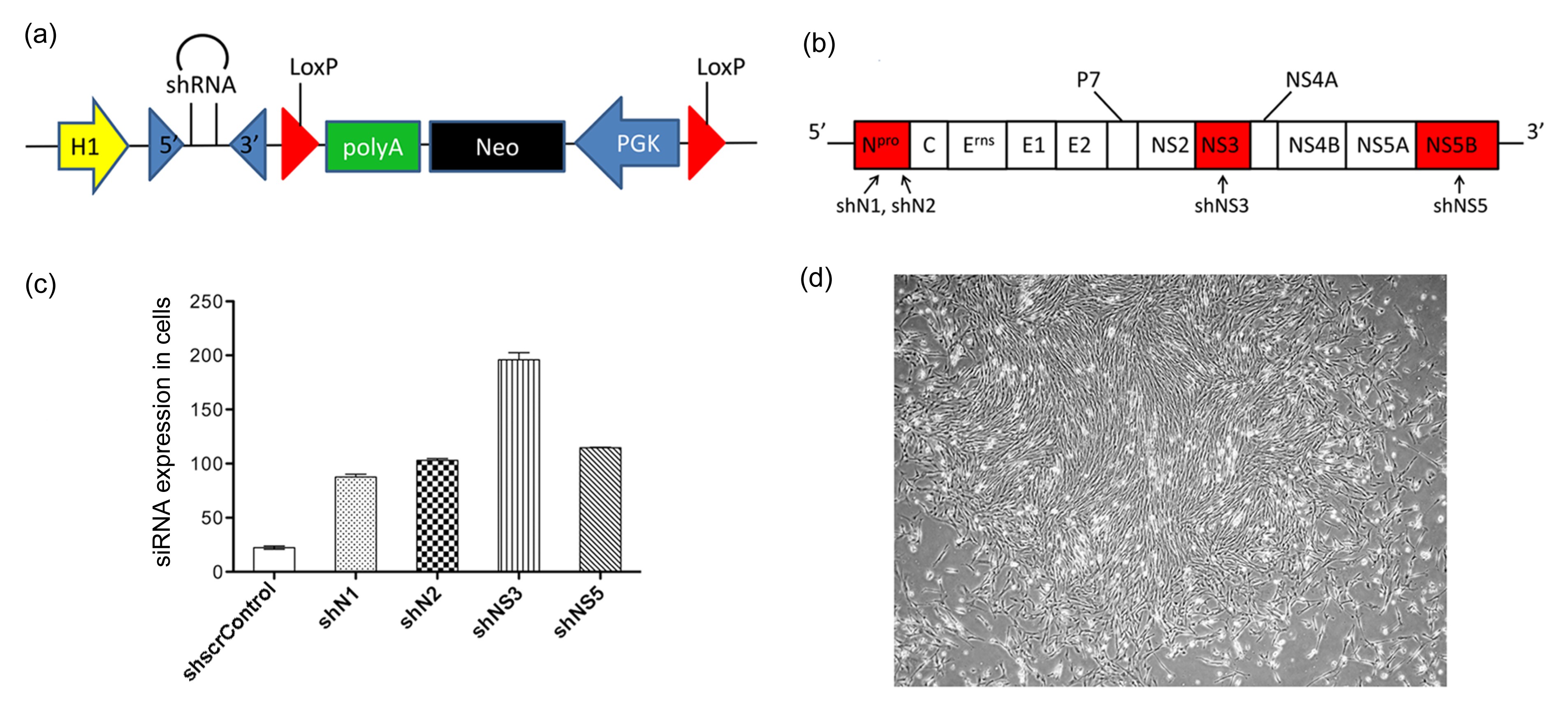
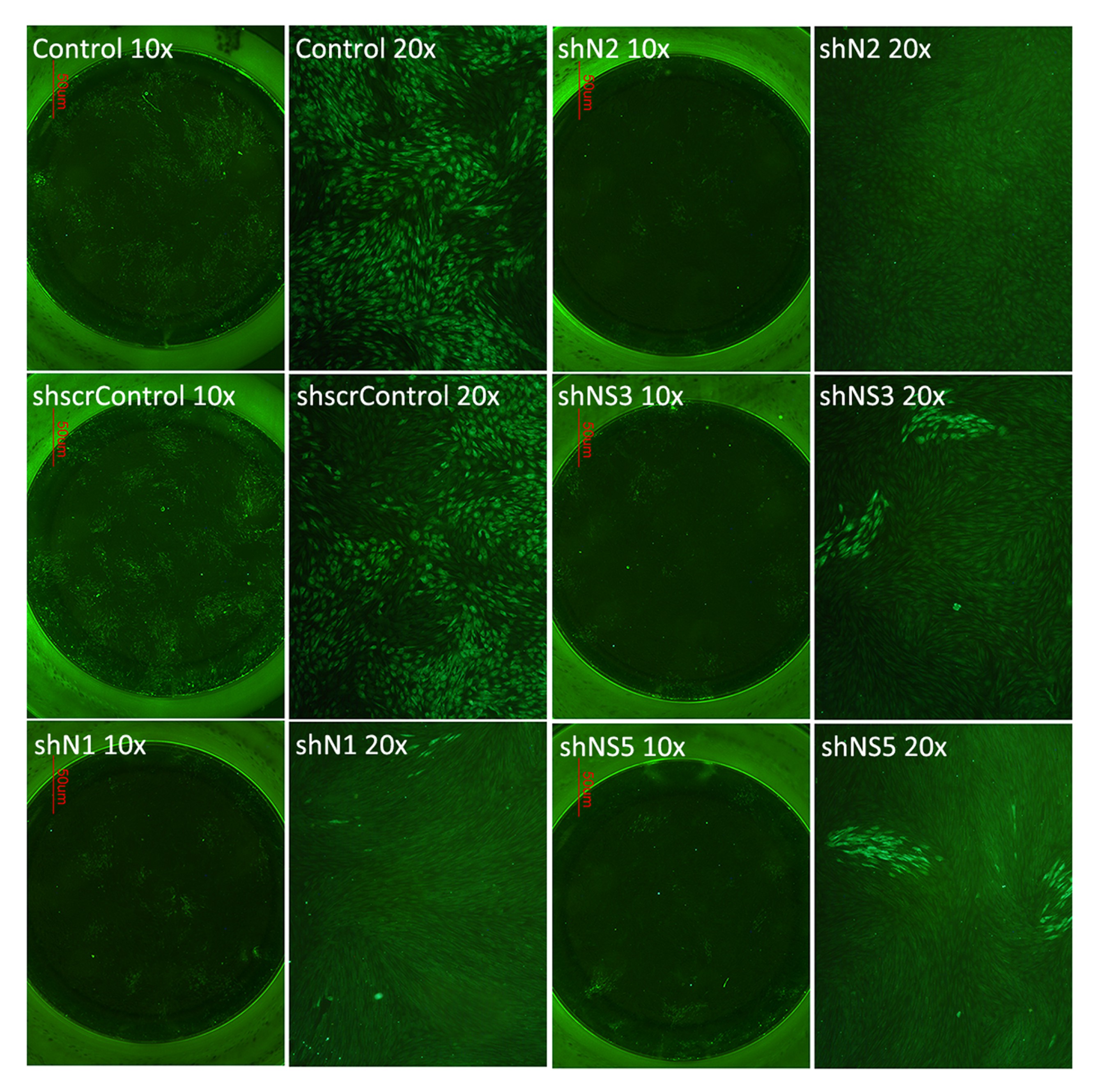
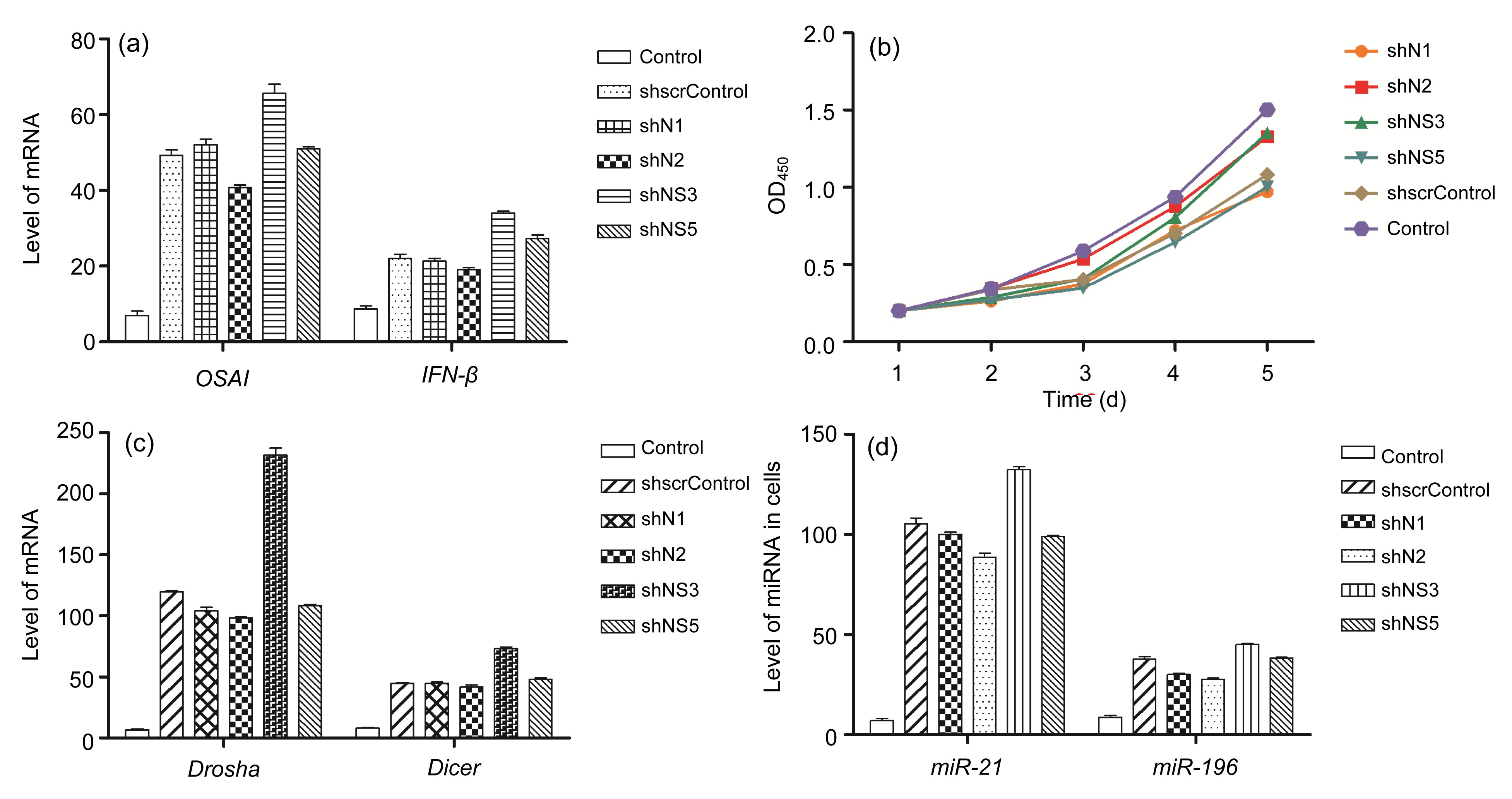
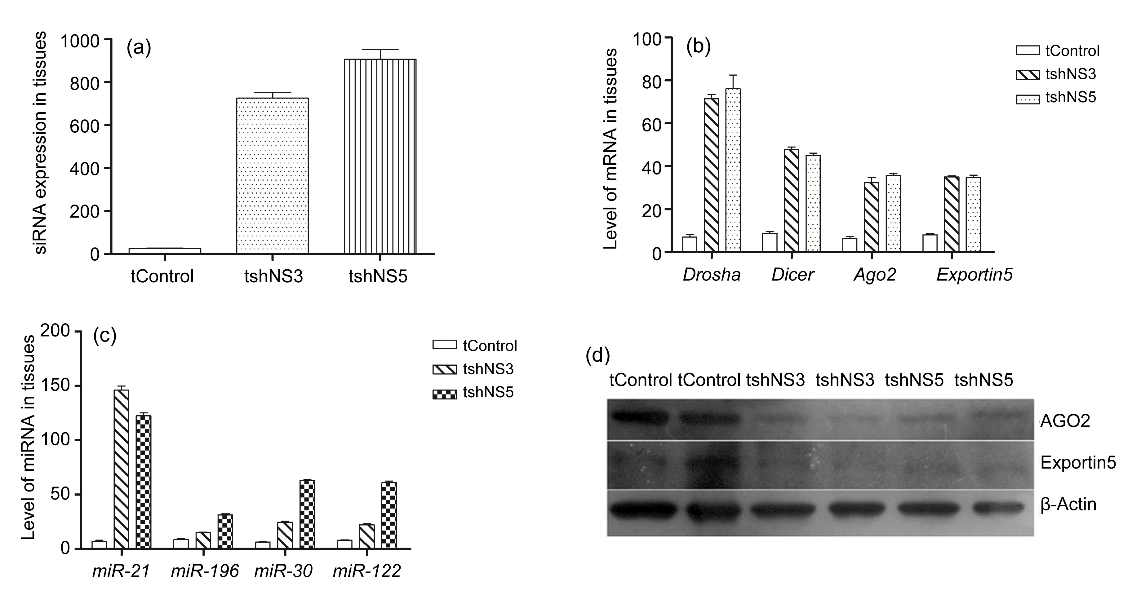
Abstract: 
RNA interference (RNAi) is considered as a potential modality for clinical treatment and anti-virus animal breeding. Here, we investigate the feasibility of inhibiting classical swine fever virus (CSFV) replication by short hairpin RNA (shRNA) in vitro and in vivo. We generate four different shRNA-positive clonal cells and two types of shRNA-transgenic pigs. CSFV could be effectively inhibited in shRNA-positive clonal cells and tail tip fibroblasts of shRNA-transgenic pigs. Unexpectedly, an early lethality due to shRNA is observed in these shRNA-transgenic pigs. With further research on shRNA-positive clonal cells and transgenic pigs, we report a great induction of interferon (IFN)-responsive genes in shRNA-positive clonal cells, altered levels of endogenous microRNAs (miRNA), and their processing enzymes in shRNA-positive cells. What is more, abnormal expressions of miRNAs and their processing enzymes are also observed in the livers of shRNA-transgenic pigs, indicating saturation of miRNA/shRNA pathways induced by shRNA. In addition, we investigate the effects of shRNAs on the development of somatic cell nuclear transfer (SCNT) embryos. These results show that shRNA causes adverse effects in vitro and in vivo and shRNA-induced disruption of the endogenous miRNA pathway may lead to the early lethality of shRNA-transgenic pigs. We firstly report abnormalities of the miRNA pathway in shRNA-transgenic animals, which may explain the early lethality of shRNA-transgenic pigs and has important implications for shRNA-transgenic animal preparation.

shRNA转基因猪中miRNA通路饱和引起的早期致死性

研究目的:通过制备抗猪瘟病毒shRNA转基因猪,探索shRNA对猪胎儿成纤维细胞、克隆猪早期胚胎发育以及转基因猪的影响。
创新要点:基于稳定表达shRNA的猪胎儿成纤维克隆细胞及shRNA转基因克隆猪制备的结果,发现shRNA对克隆猪早期囊胚发育阶段无明显毒性,并首次报道了在shRNA转基因动物中,shRNA引起体内miRNA通路过饱和及动物致死性等毒性。
研究方法:实验构建了多种抗猪瘟病毒shRNA表达载体,转染猪胎儿成纤维细胞并筛选得到细胞克隆,结合体细胞核移植技术制备了shRNA转基因猪。使用实时荧光定量聚合酶链式反应(PCR)和蛋白质印迹法(Western blotting)检测了细胞及个体水平siRNA的表达量、干扰素反应相关基因、内源miRNA及通路因子的表达量,同时检测了shRNA克隆细胞囊胚率。
重要结论:shRNA能有效抑制猪瘟病毒的复制。在猪胎儿成纤维细胞水平上,稳定表达shRNA能引起细胞内干扰素反应、miRNA通路过饱和等副作用;在克隆猪发育阶段,shRNA对克隆猪早期囊胚发育阶段无明显毒性;在shRNA转基因克隆猪个体水平上,shRNA引起克隆猪体内miRNA通路过饱和及克隆猪早期易致死等毒性。

关键词:致死性;shRNA转基因克隆猪;microRNA通路;猪瘟病毒;囊胚发育

Darkslateblue:Affiliate; Royal Blue:Author; Turquoise:Article

Article Content

1.  Introduction

 RNA interference (RNAi) is a gene silencing phenomenon which was first discovered in Caenorhabditis elegans triggered by double-stranded RNA (dsRNA) (Lecellier et al., 2005). Several researchers reported hepatotoxicity and fatality induced by ectopic RNAi triggers when they attempted to intravenously inject adeno-associated virus (AAV)-mediated shRNA vectors into mouse models (Ahn et al., 2011; Borel et al., 2011; Martin et al., 2011), which seriously hindered therapeutic RNAi. On the other hand, RNAi technology has been widely applied to inhibit viruses in vitro, including human immunodeficiency virus, hepatitis C virus, poliovirus, foot-and-mouth disease virus, porcine transmissible gastroenteritis virus, etc. (John et al., 2007; Xu et al., 2008; Lv et al., 2009). Recently, Maillard et al. (2013) demonstrated that antiviral RNAi operates in mammalian cells. However, few reports have been published regarding the production of transgenic animals resistant to viruses by RNAi. Cell reprogramming that occurs in the process of somatic cell nuclear transfer (SCNT) may convert exogenous short hairpin RNA (shRNA) cassettes into endogenous microRNAs (miRNAs). Whether in vivo toxicities induced by intravenously injected exogenous small interfering RNA (siRNA)/shRNA would occur in shRNA-transgenic animals remains controversial.

 In our study, a low survival rate and early lethality were observed in shRNA-transgenic pigs compared with other transgenic pigs when we attempted to produce shRNA-transgenic pigs with anti-CSFV (classical swine fever virus) capacity. We achieved porcine fetal fibroblasts (PFFs; large white) clones stably expressing shRNAs and produced shRNA-transgenic pigs by SCNT. Interestingly, we found that shRNAs led to the induction of interferon (IFN)-responsive genes and abnormalities in endogenous miRNAs and their processing enzymes in these clones. Saturation of the miRNA pathway and altered endogenous miRNA levels were also discovered in the transgenic pig livers, which explained the fatality of the shRNA-transgenic pigs in our experiment. Finally, we investigated the effects of shRNAs on the development of SCNT embryos by measuring the blastocyst formation rate. In conclusion, we investigated the feasibility of preparing shRNA-transgenic pigs with anti-CSFV capacity and reported early lethality of shRNA-transgenic animals caused by disruption of the endogenous miRNA pathway. Our results have a fundamental significance for the generation of shRNA-transgenic animals and antiviral RNAi in mammals.


2.  Materials and methods

2.1.  Total RNA extraction and real-time polymerase chain reaction (PCR) amplification

 RNA was extracted with TRIzol reagent (Invitrogen) and purified using an RNeasy column (Qiagen). For quantitative real-time PCR, the samples were digested with DNase I followed by the reverse transcription of 1 μg of total RNA using moloney murine leukemia virus (M-MLV; Promega) and complementary DNA (cDNA) was prepared. The target genes were amplified in three replicates using the iQtm5 multicolor real-time PCR detection system (Bio-Rad) with the BioEasy SYBR Green I real-time PCR kit (Bioer Technology, Hangzhou, China). The housekeeping gene glyceraldehyde 3-phosphate dehydrogenase (GAPDH) was selected as the internal control. The specificity of amplification was verified by evaluation of the melting curve and checking for correct size of amplification products in agarose gel. All the primers used in our research are listed in Table S1.

2.2.  Viral infections and indirect immunofluorescence assay (IFA)

 Indirect IFA was performed according to Xu et al. (2008). Positive anti-CSFV serum was obtained from the Academy of Military Medical Sciences (Changchun, China). For IFA, the cells were fixed in 80% (v/v) cold acetone for at least 30 min after being infected with CSFV for at least 50 h. The cells were then incubated with diluted positive anti-CSFV serum (1:100, v/v) at 39 °C for 1 h. The cells were washed three times by phosphate-buffered saline with Tween 20 (PBST) and incubated with a fluorescein isothiocyanate (FITC)-conjugated rabbit anti-pig IgG secondary antibody (Sigma) at 39 °C for 2 h. The cells were washed three times by PBST, and green fluorescence was examined using an ECLIPSE TE2000-V (Nikon) system.

2.3.  Western blotting and MTS assay

 The primary antibodies were Exportin5 polyclonal antibody (A01; Abnova) and Argonaute2 (AGO2) rabbit mAb (C34C6; Cell Signaling). The protein concentrations were assayed using bovine serum albumin (BSA) as the working standard. Then 30 μg proteins mixed with loading buffer were boiled before being resolved on sodium dodecyl sulfate polyacrylamide gel electrophoresis (SDS-PAGE). Next, the samples were transferred onto nitrocellulose membranes (Bio-Rad). The blots were blocked using blocking reagents (LI-COR Biosciences) for 2 h followed by incubation overnight with a primary antibody. The blots were washed three times by PBST and then incubated for 1 h in either anti-rabbit or anti-mouse IgG conjugated to horseradish peroxidase (1:15 000, w/w). Immunoreactive bands were visualized using BeyoECL Plus (Beyotime Inc., China) according to the manufacturer’s instructions. β-Actin served as a loading control.

 The cell proliferation assay was performed using cell counting kit-8 (CCK-8; Dojindo Laboratories, Kumamoto, Japan). The cells were incubated at 39 °C and at 5% CO2 for 24, 48, 72, 96, 118, or 144 h, and 10 μl of the CCK-8 solution was added. After incubating for 2 h at 39 °C, the absorbance of each well was recorded using an enzyme-linked immunosorbent assay (ELISA) plate reader at 450 nm. The analysis was performed according to Zhao et al. (2011).

2.4.  Cell culture and donor cell preparation for SCNT

 PFFs were isolated from 33-d-old fetuses and were grown in Dulbecco’s modified Eagle’s medium (DMEM; GIBCO) with 10% (v/v) fetal bovine serum (FBS) at 39 °C and 5% CO2. All the plasmids used in our research including the scrambled control, were constructed by our own laboratory. To produce stably transfected cells, the cells transfected (Fugene HD, Roche) with a linearized shRNA expression construct were dispersed into six-well dishes, and 450 ng/ml of G418 (Amresco) was added to the medium (DMEM+10% FBS) to select clones for 10–12 d. Then the surviving cell colonies were examined by PCR and IFA, and the positive cell colonies were isolated from donor cells for SCNT.

2.5.  Production of SCNT embryos and generation of transgenic pigs

 Positive cell clones were used to construct transgenic pigs by nuclear transplantation (NT). The details of SCNT embryo production were described in Lai et al. (2002). Briefly, high quality cumulus-oocyte complexes (COCs) were matured in a maturation medium for 42 to 44 h at 39 °C and 5% CO2. The cytoplasm of SCNT recipients was introduced with donor cells, and the cells were fused by 2 DC pulses of 1.2 kV/cm for 30 μs in a BTX Electro Cell Manipulator 200 (BTX, San Diego, CA, USA). Then the SCNT embryos were cultured in PZM3 for 148 h after being electrically activated. The embryos were cultured for 20 h following activation and were surgically transferred into the oviducts of surrogate pigs.


3.  Results

3.1.  Generation of PFFs and transgenic pigs stably expressing shRNAs

 To generate PFFs and shRNA-transgenic pigs with anti-CSFV capacity, we transfected PFFs with four anti-CSFV shRNA constructs (shN1, shN2, shNS3, and shNS5) and a scrambled shRNA construct (shscrControl) (Figs. 1a and 1b). After selecting by G418, 30 clones for each shRNA were identified by PCR and mixed (Fig. 1c). As shown in Fig. 1d (P<0.05), a high level of siRNAs was observed in these clones. Using IFA, we examined the level of viral antigen produced in the clones that were infected with CSFV and observed that only a few anti-CSFV shRNA cells displayed green fluorescence (Fig. 2), indicating that CSFV was effectively inhibited by shN1, shN2, shNS3, and shNS5.


Fig.1
Generation of shRNA-positive PFF clones
(a, b) siRNA expression vectors PGKneolox2-shRNA (shN1, shN2, shNS3, shNS5, and shscrControl) and genomic structure and encoding proteins of the CSFV Shimen strain; (c) Overexpressed siRNA in clonal cells compared with scrambled shRNA-positive clonal cells (shscrControl), as determined by real-time PCR (data are expressed as mean±SD, n=6; P<0.05); (d) An image of a shRNA-positive PFF clone


Fig.2
Protective effect of shRNA against CSFV infection
Viral infection in PFF clones was examined by an IFA. Only a few cells in the antiviral shRNA wells displayed green fluorescence compared with the control, indicating that the shRNA inhibits CSFV effectively

 Two types of shRNA-positive clones (shNS3 and shNS5) that showed strong antiviral effects, stable cells, and a homogeneous morphology, were selected as donor cells for preparing clonal pigs by SCNT. The results of PCR detection of the neomycin resistance gene from tail DNA of clonal pigs were all positive (data not shown). Overexpressed siRNAs in the livers of transgenic pigs were observed by real-time PCR (Fig. 4a). To detect CSFV replication, adherent cells from the tails of transgenic pigs were isolated and cultured. Our results showed that most of the cells were effectively protected by shRNA and resisted viral infection (Fig. S1).

3.2.  Adverse effects in shRNA-positive clones

 A high expression level of shRNA is considered one of the factors that cause an IFN response (Stewart et al., 2008). As expected, a 10–70-fold induction of OASI and IFN-β, two IFN-stimulated genes, was observed in the shRNA-positive cells (P<0.05) (Fig. 3a). Next, these clonal cells and non-transfected control cells were selected for an MTS assay. The PFF clones that were stably transfected with shRNAs exhibited slightly decreased proliferation compared to the non-transfected control cells (Fig. 3b).


Fig.3
Adverse effects induced by shRNA in PFFs
(a) IFN responsive genes (OASI, IFN-β) in clones increased compared with non-transfected control (P<0.05); (b) shRNA clones exhibited a little decreased proliferation compared to the non-transfected control cells; (c) miRNA processing enzyme Dicer and Drosha mRNA increased 50–250-fold in shRNA-positive cells (P<0.05); (d) Mature miRNAs including miR-21 and miR-196 in clonal cells increased significantly compared with non-transfected control (P<0.05). Data are expressed as mean±SD (n=6)

 To assess whether overexpressing shRNAs do dysfunctional miRNA pathways in vitro, we measured the mRNA levels of Dicer and Drosha, two processing enzymes in the miRNA/shRNA pathway. As shown in Fig. 3c (P<0.05), Dicer and Drosha mRNAs increased 50–250-fold in these clones. At the same time, two ubiquitous mature miRNAs, miR-21 and miR-196, increased 30–150-fold (Fig. 3d) (P<0.05), which also demonstrated the disruption of the endogenous miRNA pathway.

3.3.  Abnormalities of endogenous miRNA pathway in shRNA-transgenic pigs

 To gain insight into RNAi-related toxicities in vivo, two types of shRNA-transgenic pigs (shNS3 and shNS5) were selected for our subsequent studies. We collected the livers of four shNS3-transgenic pigs and four shNS5-transgenic pigs, which all died about one week old. Similar to shRNA clones, the mRNA levels of miRNA processing enzymes in the livers of transgenic pigs increased strongly (P<0.05; Fig. 4b). More types of ubiquitous mature miRNAs, including miR-21, miR-30, miR-122, and miR-196, were tested in the livers of transgenic pigs, and all miRNAs showed an increasing trend in expression compared to the control (P<0.05; Fig. 4c).


Fig.4
Abnormalities of miRNA pathway in the livers of transgenic pigs
(a) Overexpressed siRNA in transgenic animal liver tissue compared with normal liver tissue (tControl), as determined by real-time PCR (P<0.05); (b) All the miRNA processing enzymes expressed at a high level in the liver tissue of transgenic pigs (P<0.05); (c) More endogenous ubiquitous miRNAs, including miR-21, miR-30, miR-122, and miR-196, were detected and showed an increasing expression in livers of shRNA transgenic pigs (P<0.05); (d) Western blotting analysis of the AGO2 protein and Exportin5 protein in liver tissue. AGO2 decreased sharply in tissue samples in two replicates compared with the control (tControl), while Exportin5 decreased slightly. Data are expressed as mean±SD (n=6)

 Interestingly, the level of AGO2 protein was inversely correlated with the overexpressed shRNA, as determined by Western blotting, and Exportin5 protein expression was reduced weakly compared with the control (Fig. 4d). These results demonstrated that shRNA-transgenic animals also show RNAi-related toxicities, although cell reprogramming may convert exogenous shRNAs into endogenous miRNAs in transgenic pigs after the SCNT process.

3.4.  Blastocyst formation rate for donor cells stably transfected with shRNAs

 Several studies have demonstrated that injection of siRNA could cause non-specific abnormal development in zebrafish and abnormal blastocyst formation (Adachi et al., 2007; Zhao et al., 2008). For producing cloned embryos, four types of anti-CSFV shRNA-positive clonal cells, scrambled shRNA clonal cells, and normal control cells were transferred into enucleated oocyte recipient cells at the metaphase II stage. Then, the SCNT embryos were cultured in PZM3 for 148 h. The development of SCNT embryos was evaluated by measuring the cleavage and blastocyst formation rates (Huang et al., 2011). In our experiment, the rate of blastocyst formation did not show significant difference between the experimental and control groups (Table 1), suggesting that adverse effects caused by shRNA may occur in later developmental stages.


Table 1

Statistics of nuclear transfer blastocyst formation rate
Group No. of embryos No. (percent) of cleavages
No. (percent) of blastocysts
24 h 48 h
Control 160 87 (54.38%)a 140 (87.50%)a 20 (12.50%)a
shscrControl 162 85 (52.47%)a 135 (83.33%)b 20 (12.35%)a
shN1 148 90 (60.81%)b 132 (89.19%)a 16 (10.81%)a
shN2 152 80 (52.63%)a 140 (92.10%)b 20 (13.16%)a
shNS3 155 93 (60.00%)b 137 (88.39%)a 10 (6.45%)b
shNS5 158 84 (53.16%)a 134 (84.81%)b 13 (8.23%)b

  • All experimental groups are compared to the control. a, b Values with different superscripts within a column are significantly different


  • 4.  Discussion

     In our previous work, a low survival rate and early lethality were observed in shRNA-transgenic pigs compared with other transgenic pigs (data not shown). Cao et al. (2005) reported the phenomenon that DNA constructs designed to produce short hairpin, interfering RNAs in transgenic mice sometimes show early lethality and an IFN response. To confirm these findings, Grimm et al. (2006) intravenously injected 49 AAV/shRNA vectors into mice and identified hepatotoxicity and fatality in the mice which are related to exogenous siRNA/shRNA concentrations rather than off-targets. However, cell reprogramming that occurs in the process of SCNT may convert exogenous shRNA cassettes into endogenous miRNAs. Whether in vivo toxicities induced by intravenously injected exogenous siRNA/shRNA would occur in shRNA-transgenic animals remains controversial.

     In our study, we achieved shRNA-positive PFFs and livers of shRNA-transgenic pigs which died at approximately one week old. All shRNAs were designed with low homology to swine; thus, the early lethality appeared to be unrelated to off-target effects. Our results showed that all the anti-CSFV shRNAs have antiviral capacity in both PFFs clones and tail fibroblasts from transgenic pigs stably expressing shRNAs. shN1, shN2, shNS3, shNS5, and the scrambled control all triggered the induction of IFN-responsive genes in these clonal cells, indicating that induction was sequence-independent. Many studies have claimed that unmethylated CpG motifs in many vectors may also induce the IFN effect in vitro and in vivo. However, this effect is unlikely to explain the induction of IFN in clonal cells because the CpG motifs in vectors are easily de novo methylated when stably integrated into the genome (Stewart et al., 2008). Previous research has indicated that sequences of short siRNAs with a 5′ phosphate group can also trigger an endogenous IFN response (Bridge et al., 2003; Kim et al., 2004), which agrees with the IFN effect we observed.

     To assess whether fatalities in these shRNA-transgenic pigs are related to disruption of miRNA pathways, we studied the expression of several ubiquitous endogenous mature miRNAs and found abnormal expression of endogenous miRNA due to shRNA expression in vitro and in vivo. Furthermore, abnormality of miRNAs may disorder related genes and other toxicities (Yuan et al., 2014). Lecellier et al. (2005) discovered that miR-196 is related to development and can regulate the HoxB8 gene in mice. These results showed that exogenous shRNAs affect endogenous mature miRNA levels not only in primary cell clones but also in shRNA transgenic animals, which may be another explanation for RNAi-relative toxicities.

     Lethal toxicity would be observed in the early development of embryonic stem cells due to a lack of the Dicer enzyme (Kuehbacher et al., 2007). In our study, miRNA processing enzymes in both shRNA clonal cells and transgenic animals exhibit a significant rise compared with control, providing an additional option for an adverse effect caused by shRNA. Furthermore, high levels of mRNA expression of these enzymes also remind us that protein translation hysteresis is occurring. Ago2 and Exportin5 have been thought to be a rate-limiting determinant for RNAi efficacy (Grimm et al., 2006). Our data showed a large variation in AGO2 levels due to competitions between shRNAs and miRNAs. Interestingly, the levels of the Exportin5 protein did not vary noticeably, which may result from Exportin5’s role as a nucleoporin. These results corroborate the study of Martin et al. (2011) in therapeutic RNAi. Thus, we may explain the fatalities in shRNA-transgenic pigs by the fact that the shRNA induced abnormal expression of endogenous miRNAs and its processing enzymes.

     Recently, Zhao et al. (2008) found that RNAi could cause non-specific abnormal development in zebrafish, as siRNA injection caused a low level of miR-430 expression. Adachi et al. (2007) found that abnormal blastocyst formation and trophectoderm differentiation defects were caused by injecting siRNAs. In previous studies, siRNAs have always targeted endogenous genes, while we aimed to use shRNAs to inhibit exogenous CSFV. Here we assume that adverse effects caused by shRNA may occur in later developmental stages because the shRNA does not cause an obvious effect on blastocyst development.


    5.  Conclusions

     In summary, our findings showed that shRNA caused adverse effects in vitro and in vivo, and shRNA-induced disruption of endogenous miRNA pathway may lead to the early lethality of shRNA-transgenic pigs. Further studies need to be performed to understand the detailed mechanisms of antiviral RNAi in mammals and the preparation of shRNA-transgenic animals.


    * Project supported by the Major Projects of New Varieties of Genetically Modified Organisms (No. 2011ZX08006-001) and the Program for Changjiang Scholars and Innovative Research Team in the University (No. IRT1248), China# Electronic supplementary materials: The online version of this article (http://dx.doi.org/10.1631/jzus.B1400001) contains supplementary materials, which are available to authorized usersCompliance with ethics guidelines Zhen DAI, Rong WU, Yi-cheng ZHAO, Kan-kan WANG, Yong-ye HUANG, Xin YANG, Zi-cong XIE, Chang-chun TU, Hong-sheng OUYANG, Tie-dong WANG, and Da-xin PANG declare that they have no conflict of interest.


    References

    [1] Adachi, K., Soeta-Saneyoshi, C., Sagara, H., 2007. Crucial role of Bysl in mammalian preimplantation development as an integral factor for 40S ribosome biogenesis. Mol Cell Biol, 27(6):2202-2214. 


    [2] Ahn, M., Witting, S.R., Ruiz, R., 2011. Constitutive expression of short hairpin RNA in vivo triggers buildup of mature hairpin molecules. Human Gene Therapy, 22(12):1483-1497. 


    [3] Borel, F., van Logtenstein, R., Koornneef, A., 2011.  In vivo knock-down of multidrug resistance transporters ABCC1 and ABCC2 by AAV-delivered shRNAs and by artificial miRNAs. J RNAi Gene Silencing, 7:434-442. 


    [4] Bridge, A.J., Pebernard, S., Ducraux, A., 2003. Induction of an interferon response by RNAi vectors in mammalian cells. Nat Genet, 34(3):263-264. 


    [5] Cao, W., Hunter, R., Strnatka, D., 2005. DNA constructs designed to produce short hairpin, interfering RNAs in transgenic mice sometimes show early lethality and an interferon response. J Appl Genet, 46(2):217-225. 


    [6] Grimm, D., Streetz, K.L., Jopling, C.L., 2006. Fatality in mice due to oversaturation of cellular microRNA/short hairpin RNA pathways. Nature, 441(7092):537-541. 


    [7] Huang, Y.Y., Tang, X.C., Xie, W.H., 2011. Vitamin C enhances in vitro and in vivo development of porcine somatic cell nuclear transfer embryos. Biochem Biophys Res Commun, 411(2):397-401. 


    [8] John, M., Constien, R., Akinc, A., 2007. Effective RNAi-mediated gene silencing without interruption of the endogenous microRNA pathway. Nature, 449(7163):745-747. 


    [9] Kim, D.H., Longo, M., Han, Y., 2004. Interferon induction by siRNAs and ssRNAs synthesized by phage polymerase. Nat Biotechnol, 22(3):321-325. 


    [10] Kuehbacher, A., Urbich, C., Zeiher, A.M., 2007. Role of Dicer and Drosha for endothelial microRNA expression and angiogenesis. Circ Res, 101(1):59-68. 


    [11] Lai, L., Park, K.W., Cheong, H.T., 2002. Transgenic pig expressing the enhanced green fluorescent protein produced by nuclear transfer using colchicine-treated fibroblasts as donor cells. Mol Reprod Dev, 62(3):300-306. 


    [12] Lecellier, C.H., Dunoyer, P., Arar, K., 2005. A cellular microRNA mediates antiviral defense in human cells. Science, 308(5721):557-560. 


    [13] Lv, K., Guo, Y., Zhang, Y., 2009. Transient inhibition of foot-and-mouth disease virus replication by siRNAs silencing VP1 protein coding region. Res Vet Sci, 86(3):443-452. 


    [14] Maillard, P.V., Ciaudo, C., Marchais, A., 2013. Antiviral RNA interference in mammalian cells. Science, 342(6155):235-238. 


    [15] Martin, J.N., Wolken, N., Brown, T., 2011. Lethal toxicity caused by expression of shRNA in the mouse striatum: implications for therapeutic design. Gene Therapy, 18(7):666-673. 


    [16] Stewart, C.K., Li, J., Golovan, S.P., 2008. Adverse effects induced by short hairpin RNA expression in porcine fetal fibroblasts. Biochem Biophys Res Commun, 370(1):113-117. 


    [17] Xu, X., Guo, H., Xiao, C., 2008.  In vitro inhibition of classical swine fever virus replication by siRNAs targeting Npro and NS5B genes. Antiviral Res, 78(3):188-193. 


    [18] Yuan, Z.M., Yang, Z.L., Zheng, Q., 2014. Deregulation of microRNA expression in thyroid tumors. J Zhejiang Univ-Sci B (Biomed & Biotechnol), 15(3):212-224. 


    [19] Zhao, X.F., Fjose, A., Larsen, N., 2008. Treatment with small interfering RNA affects the microRNA pathway and causes unspecific defects in zebrafish embryos. FEBS J, 275(9):2177-2184. 


    [20] Zhao, Y., Pang, D., Wang, T., 2011. Human MxA protein inhibits the replication of classical swine fever virus. Virus Res, 156(1-2):151-155. 


    Open peer comments: Debate/Discuss/Question/Opinion

    <1>

    Please provide your name, email address and a comment


    Journal of Zhejiang University-SCIENCE, 38 Zheda Road, Hangzhou 310027, China
    Tel: +86-571-87952783; E-mail: cjzhang@zju.edu.cn
    Copyright © 2000 - 2024 Journal of Zhejiang University-SCIENCE