1. Introduction
Oxytetracycline (OTC) is a broad spectrum antibiotic in the tetracycline (TC) group. Its chemical formula is C
22H
24N
2O
9 with a molecular weight of 460.4 Da. It is authorized for use in medicated feed in aquaculture (Serrano,
2005) and is usually administered through the feed during the growing period with a recommended withdrawal period of 4–16 d before harvesting, depending on the concentration, water temperature, and shrimp species (Wang et al.,
2004; Nogueira-Lima et al.,
2006; Gómez-Jimenez et al.,
2008). Overuse and insufficient lengths of withdrawal periods lead to OTC residues in aquaculture products, causing a serious threat to human health. TCs, including OTC, have an impact on human health in two different ways: at high concentrations, they inhibit mammalian protein synthesis, while at low concentrations, they cause acquired resistance in microorganisms (Srisomboon and Poomchatra,
1995).
To prevent harmful health effects in consumers due to OTC residues, maximum residual limits (MRLs) have been established for OTC. The European Union (EU) proposed MRLs of 100 ng/g for muscle, 300 ng/g for liver, and 600 ng/g for kidney for all animals used for human consumption (The Council of the European Communities,
1990). These limits require sensitive and specific methods for the detection of OTC in food-producing animals. Various methods have been reported, most of which are based on chemical methods such as high-performance liquid chromatography (HPLC) (Furusawa,
1999; Cinquina et al.,
2003; Yuan et al.,
2010), liquid chromatography-mass spectroscopy (LC-MS) (Carson et al.,
1998; Cherlet et al.,
2003), and capillary electrophoresis (Huang et al.,
1997). Although these methods offer a high precision and accuracy, they require expensive equipment and well-trained professionals. Moreover, most chemical methods require a complicated sample preparation step. Commonly, cleanup methods are based on solid-phase extraction (Furusawa,
1999; Cherlet et al.,
2003; Cliquet et al.,
2003) or metal chelate affinity chromatography (Croubels et al.,
1997). These methods are time-consuming and costly, and require large sample volumes.
The enzyme-linked immunosorbent assay (ELISA), an immunological method based on the specific binding between an antibody and an antigen, has become a method of choice for routine screening because of its high sensitivity, simplicity, cost effectiveness, and ability to screen large numbers of samples in a short period. Importantly, an immunoassay does not require a complicated step to clean up samples (Cháfer-Pericása et al.,
2010). Previously, established ELISA methods have been based on a polyclonal antibody (PAb) for doxycycline (DC) detection (Le et al.,
2009) and a monoclonal antibody (MAb) for chlortetracycline (CTC) detection (Le et al.,
2011b). The use of a MAb is preferable due to its uniformity and unlimited production with unchanged properties. Nevertheless, the production of a highly sensitive MAb for the detection of OTC has not yet been reported.
In this study, the generation of a MAb specific for OTC, the detection of OTC residues in shrimp samples using ELISA, and a simple sample preparation method for routine detection are reported.
2. Materials and methods
2.1. Materials, reagents, and animals
Ninety-six-well ELISA and culture plates were purchased from Corning Incorporated (NY, USA). OTC hydrochloride and other TC antibiotics, bovine serum albumin (BSA), ovalbumin (OVA), complete and incomplete Freund’s adjuvants, polyethylene glycol (PEG) (molecular weight 3000–3700), 3,3',5,5'-tetramethylbenzidine (TMB), and ethylenediaminetetraacetic acid (EDTA) tetrasodium salt dihydrate were purchased from the Sigma Chemical Co. (MO, USA). Hybridoma serum-free medium was purchased from GIBCO (NY, USA). Fetal bovine serum (FBS) was purchased from PAA Laboratories (Austria). Goat anti-mouse IgG-horseradish peroxidase (HRP) was purchased from Jackson Immuno Research Laboratories. Female BALB/c mice aged between 8 and 10 weeks were purchased from the National Laboratory Animal Centre (Mahidol University, Salaya, Thailand). All procedures involving laboratory animals were conducted in accordance with guidelines and approved by the Institutional Animal Care and Use Committee, Institute of Biotechnology and Genetic Engineering, Chulalongkorn University, Thailand.
2.2. Preparation of OTC-carrier protein conjugates
OTC was conjugated to a carrier protein (BSA for immunization and OVA for ELISA) using the Mannich reaction (Faraj and Ali,
1981). Four milligrams of OTCs were dissolved in 0.2 ml of 30% ethanol and mixed with 10 mg of carrier protein in 0.3 ml of distilled water. A mixture of 3.0 mol/L sodium acetate buffer, pH 5.5 (0.2 ml), and 7.5% formaldehyde (0.2 ml) was added and stirred overnight at room temperature. The mixture was dialyzed against 0.01 mol/L phosphate buffer saline (PBS), pH 7.4, five times. After the dialysis, the conjugated OTC was filtered through a 0.2-μm cellulose acetate membrane and kept at −20 °C until used.
2.3. Immunization of mice
Female BALB/c mice were immunized by intraperitoneal (ip) injection with 100 μg of OTC-BSA in 50 μl of sterile PBS emulsified with 50 μl of Freund’s complete adjuvant. After the initial injection, the mice were boosted at two-week intervals with the same antigen but mixed with Freund’s incomplete adjuvant. One week after the fourth boost, the mice were tail-bled and anti-serum was collected. The antibody titer was determined using indirect ELISA (iELISA), and the antibody affinity for free OTC was determined using indirect competitive ELISA (icELISA) with OTC-OVA as the coating agent. The mice were finally boosted with 100 μg of OTC-BSA in normal saline 4 d before cell fusion.
2.4. Production of hybridomas
The fusion protocol was modified from that of Harlow and Lane (
1988). Briefly, the immunized mice were sacrificed, and their splenocytes fused with P3/NS-1/1-Ag4-1 mouse myeloma cells using 50% PEG as the fusion reagent. The fused cells were re-suspended in selective medium (hypoxanthine-aminopterin-thymidine (HAT) medium containing 20% FBS) and distributed into 96-well tissue culture plates. The hybridomas were cultured at 37 °C and 5% CO
2 with a suitable medium replacement period. About 10 to 14 d after fusion, culture supernatants were screened for antibodies using iELISA. Antibodies from the positive wells were then tested for their ability to bind free OTC using icELISA. The cultures secreting specific antibodies were cloned by multiple rounds of limiting dilution culture until monoclones were obtained.
2.5. ELISA procedures
iELISA was used for screening the secreted antibodies against OTC. For this assay, 96-well plates were coated with 5 μg/ml OTC-OVA in PBS (100 μl/well) by incubation at 4 °C overnight. The plates were washed three times with washing buffer (PBS containing 0.05% Tween-20; PBST) and blocked with blocking solution (0.05 g/ml skim milk powder in PBS, 300 μl/well) by incubation at 37 °C for 1 h. After three washes, the plates were coated with primary antibody (mouse sera or culture supernatant, 100 μl/well) at 37 °C for 2 h. Subsequently, the plates were washed again and incubated with secondary antibody (1:10 000 (v/v) goat anti-mouse IgG-HRP in PBS, 100 μl/well) at 37 °C for 1 h. After another washing, 100 ml of a substrate solution containing 0.25 g/L TMB in 205 mmol/L potassium citrate buffer, pH 4.0, and 3.0 mmol/L H
2O
2 (Frey et al.,
2000) was added. The reaction was allowed to occur in the dark for 15 min at room temperature and was then stopped with 100 μl/well of 1.0 mol/L H
2SO
4. Absorbance at 450 nm was measured using a microplate reader.
icELISA was used to screen for specific antibodies against free OTC. The procedure for icELISA was the same as that used for iELISA with the exception that the primary antibody was added together with 100 μl/well free OTC as a competitor.
2.6. Determination of MAb isotypes
Isotypes of the selected MAbs were determined using an isotyping kit (Sigma-Aldrich) according to the manufacturer’s instructions. Briefly, a 96-well plate was coated with 100 μl/well of specific antibodies against different isotypes of mouse antibody (IgG
1, IgG
2a, IgG
2b, IgG
3, IgA, and IgM) at a dilution of 1:1000 (v/v) in PBS and incubated at 4 °C for 1 h afterwards, and the plate was washed three times with PBST and blocked with skimmed milk (300 μl/well) by incubation at 37 °C for 1 h. After washing three times, 100 μl/well of a culture medium sample was added to each well, and the plate was incubated at 37 °C for 1 h. The plate was washed again and incubated with a 1:2000 (v/v) dilution of an HRP-rabbit anti-mouse Fab-specific antibody in the dark at room temperature for 30 min. After another washing, the enzyme assay was conducted as described above.
2.7. Production and purification of MAb
The selected monoclone was cultured in a 500-ml spinner flask containing serum-free medium. MAb in the hybridoma culture medium was purified by affinity chromatography using Protein G Sepharose 4 Fast Flow (Pharmacia Biotech AB, Uppsala, Sweden). The protein G column was equilibrated with 2.0 mmol/L PBS, pH 7.0, at a flow rate of 1.0 ml/min. Hybridoma culture medium (500 ml) was loaded into the column, and then unbound protein was washed out using the equilibrated buffer. MAb was eluted with 0.1 mol/L glycine-HCl buffer, pH 2.7, and collected fractionally for 30 fractions (1 ml each) in 70 μl of 1.0 mol/L Tris-HCl buffer, pH 9.0, to neutralize the pH. The protein concentration of each fraction was determined by spectrometry at 280 nm. Subsequently, fractions with high protein concentrations were pooled and dialyzed against PBS five times. The MAb was then filtered through a 0.2-μm cellulose acetate membrane and kept at −20 °C until used. The concentration of the MAb was determined using a BCA protein kit (Pierce, Rockford, IL, USA).
2.8. Determination of sensitivity and specificity of MAbs
To evaluate the sensitivity and specificity of the selected MAbs, icELISA was performed as described previously. In addition to OTC, other antibiotics of interest were tested as competitors.
The sensitivities of the MAbs were reported in terms of 50% inhibitory concentration (IC
50) and limit of detection (LOD). An IC
50 was defined as the concentration at which 50%
B/
B
0 was obtained, where
B and
B
0 are the averages of the absorbances obtained from icELISAs with and without different concentrations of OTC, respectively. The LOD was the OTC concentration of the mean blank determination signal (
B
0) reduced by three times its standard deviation (SD) (Cliquet et al.,
2003; Le et al.,
2003).
The specificities were evaluated in terms of cross-reactivity with other antibiotics. The IC
50 values of the compounds of interest were obtained as previously described. The percentage of cross-reactivity was then calculated using the following formula:
 = }}\frac{{{{\text{IC}}_{50}}{\text{ of OTC}}}}{{{{\text{IC}}_{50}}{\text{ of competitor}}}} \times {\text{100\% }}\]
)
.
2.9. Preparation of shrimp samples
One-gram samples of homogenized shrimp tissue were fortified with OTC at final concentrations of 25, 50, 100, 200, and 400 ng/g. Each sample was extracted with a mixture of 0.1 mol/L EDTA (100 μl), methanol (1 ml), and acetonitrile (1 ml). Each homogenate was vortexed for 1 min and then centrifuged at 2700×
g for 10 min. After the supernatant was collected, the same extraction procedure was repeated twice, and the supernatants were combined. The combined supernatants were then evaporated under a nitrogen stream at 45 °C. The residue was re-suspended in 10 ml PBS for icELISA analysis.
3. Results and discussion
3.1. Preparation of immunogen
OTC, which has a molecular weight of 460.4 Da, is a hapten molecule and cannot trigger the immune system of animals to produce antibodies. Therefore, it must be conjugated to a carrier protein. In this study, OTC was conjugated to BSA using the Mannich reaction. The level of conjugation was assessed by comparing the mass of BSA with the mass of the conjugated product using matrix-assisted laser desorption-ionization time of flight mass spectrometry (MALDI-TOF-MS). The result indicated that the mass of the conjugate was higher than that of BSA by 3241 Da (data not shown). Therefore, the molecular ratio of OTC to BSA in the conjugate was calculated to be 7:1. This was lower than the value for conjugation of TC-BSA, previously reported at (30–40):1 (Faraj and Ali,
1981).
3.2. Production of MAbs
After BALB/c mice were immunized with the OTC-BSA conjugate, the anti-serum titer was analyzed using iELISA. The immunization was successful, with the serum titers ranging between 1:16 000 and 1:128 000 (Table
1). All sera were subjected to icELISA using 50 μg/ml of OTC as the competitor to determine whether the antisera could bind the free OTC molecule in addition to the OTC-BSA conjugate. These experiments showed that the amount of competition was between 19% and 58% (Table
1), indicating that the antisera could bind free OTC.
Table 1
Summary of hybridomas derived from cell fusion
| Mouse No. |
Antibody titer |
Amount of competition with free OTC (%) |
Growing hybridomas (well) |
Producing antibody (well) |
Monoclone |
| 1 |
1:64 000 |
28 |
768 |
18 |
– |
| 2 |
1:64 000 |
49 |
1152 |
72 |
– |
| 3 |
1:128 000 |
35 |
768 |
8 |
– |
| 4 |
1:32 000 |
44 |
1152 |
82 |
7-3G |
| 5 |
1:128 000 |
19 |
716 |
5 |
– |
| 6 |
1:64 000 |
45 |
784 |
4 |
– |
| 7 |
1:128 000 |
58 |
960 |
9 |
– |
| 8 |
1:16 000 |
50 |
480 |
2 |
– |
| 9 |
1:32 000 |
43 |
960 |
7 |
2-4F |
| 10 |
1:64 000 |
52 |
960 |
15 |
11-11A |
In the hybridoma cell preparation, 222 of the total number of cultured wells (8700) were found to produce antibodies that bound to the OTC-BSA conjugate (Table
1). The cell culture supernatants from these wells were then tested using an icELISA with OTC as the competitor. Only three cultures showed the ability to produce antibodies with free-OTC-binding ability. These cells were re-cloned and subcloned by limiting dilution until the monoclones 2-4F, 7-3G, and 11-11A were obtained.
Most of the reported antibodies specific for TC group antibiotics are PAbs (Zhao et al.,
2008; Le et al.,
2009; Cháfer-Pericása et al.,
2010). Because the properties of PAbs vary among batches according to the immune responses of different immunized animals, PAbs are less suitable for long-term application than MAbs (Hock et al.,
1995). In addition, MAbs against CTC and DC have been reported (Le et al.,
2011a;
2011b). However, these MAbs had low cross-reactivity to OTC.
3.3. Characterization of MAbs
Isotypes of the MAbs obtained were identified using a commercial isotyping kit based on a sandwich ELISA with Fc- and Fab-specific antibodies. The isotypes of MAbs 2-4F and 11-11A were found to be IgG
1, while that of MAb 7-3G was found to be IgG
2a. The MAbs were used in an icELISA to determine their detection sensitivities in terms of their IC
50 and LOD values (Fig.
1). The results showed that MAb 2-4F was the most sensitive clone, with an IC
50 of 7.01 ng/ml and an LOD of 0.69 ng/ml, which was lower than the MRL values enforced. In contrast, the LODs of MAbs 7-3G and 11-11A were found to be 200 and 960 ng/ml, respectively, which were higher than the MRL value. Consequently, MAb 2-4F was selected for large-scale antibody production in a spinner flask. After purification using a Protein G Sepharose affinity column, the molecular weights of the heavy and the light chains of MAb 2-4F were analyzed by sodium dodecyl sulfate polyacrylamide gel electrophoresis (SDS-PAGE) and found to be 59 and 26 kDa, respectively (data not shown), in agreement with the values previously reported (Howard and Kaser,
2007).
Fig.1
MAbs from three different clones in icELISA using OTC-OVA as the coating antigen and OTC as the competitor
3.4. Sensitivity and specificity of MAb 2-4F
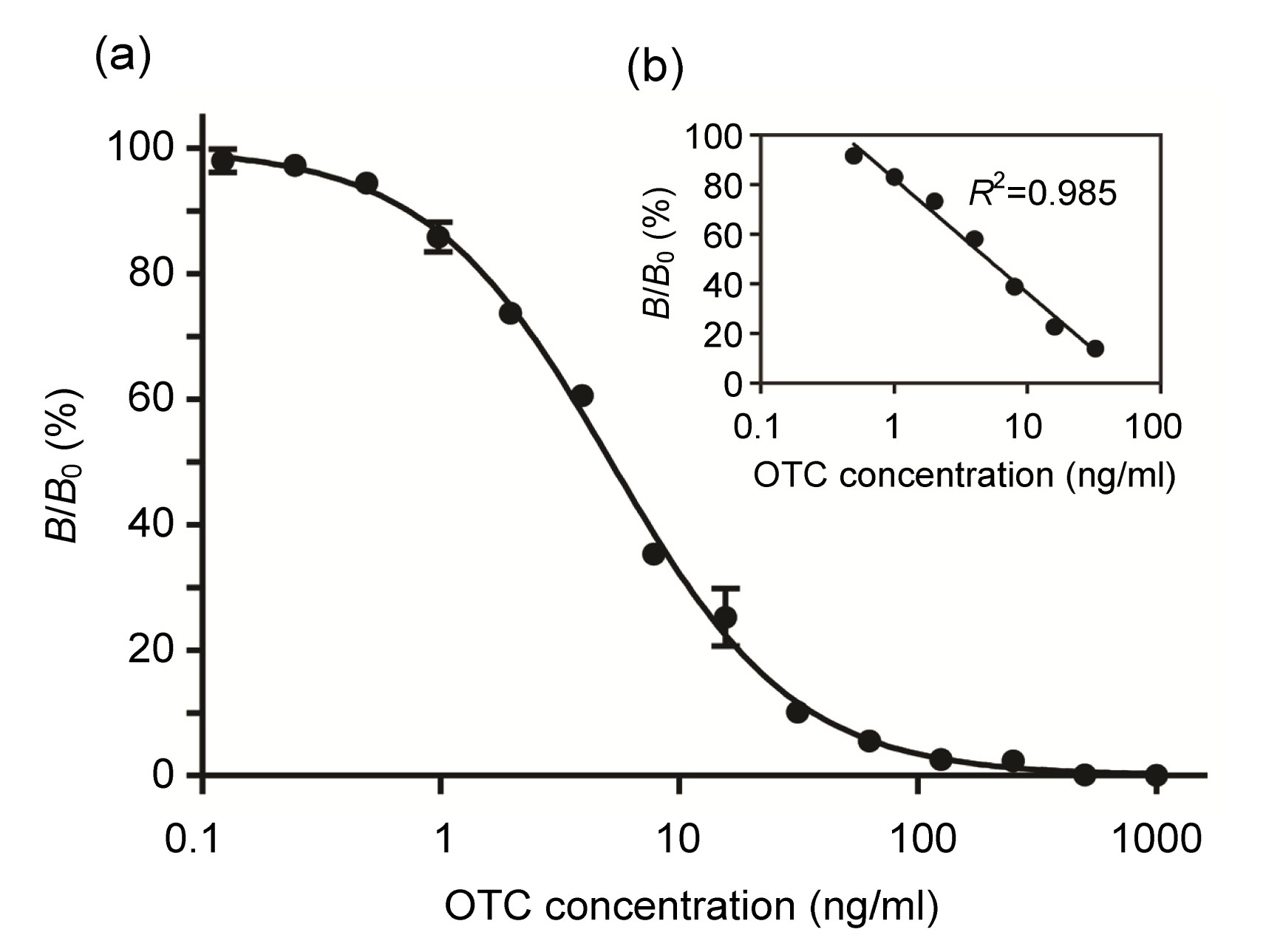
The partially purified MAb 2-4F was used in an icELISA to determine its detection sensitivity in terms of its IC
50 and LOD. A typical competitive binding curve is shown in Fig.
2a. The IC
50 and LOD were 5.16 ng/ml and 0.52 ng/ml, respectively, which were slightly lower than those obtained before purification. The linear range of the curve between 0.50 and 32.0 ng/ml exhibited an
R
2 of 0.985 (Fig.
2b).
Fig.2
Competitive inhibition curve for OTC detection by icELISA (a) and the linear range of the standard curve for OTC residue detection (b)
The assay was carried out in triplicate (mean±SD) using 2 μg/ml OTC-OVA as the coating antigen and 0.03 μg/ml MAb 2-4F
The specificity of the MAb was also determined in terms of its cross-reactivity against several compounds (Table
2). MAb 2-4F showed high cross-reactivity with rolitetracycline (RTC) (388%) but low cross-reactivity with other TCs and tested chemicals. A PAb produced against OTC had previously been reported, which also showed high cross-reactivity with TC (1028%) and RTC (449%) (Cháfer-Pericása et al.,
2010).
Table 2
Cross-reactivity of anti-OTC MAb 2-4F
| Compound |
IC50 (ng/ml) |
Cross-reactivity (%) |
| Oxytetracycline (OTC) |
5.16 |
100 |
| Rolitetracycline (RTC) |
1.33 |
388 |
| Tetracycline (TC) |
138 |
3.73 |
| Chlortetracycline (CTC) |
575 |
0.90 |
| Doxycycline (DC) |
1639 |
0.31 |
| Chloramphenicol |
>3000 |
<0.2 |
| Norfloxacin |
>3000 |
<0.2 |
| 3-Amino-5-morpholinomethyl-2-oxazolidinone (AMOZ) |
>3000 |
<0.2 |
3.5. OTC analysis in fortified shrimp samples
Minced fresh shrimp samples were spiked with known amounts of OTC at final concentrations ranging from 25 to 400 ng/g in order to cover the MRL value currently enforced at 100 ng/ml. The samples were carefully extracted because OTC binds strongly to proteins and interacts with many ions to form stable complexes (Yuwono and Indrayanto,
2005). The amount of OTC in the extracts was quantified using icELISA. The intra-assay variation was assessed as the average of six replicated wells in one microplate of each concentration sample, and the inter-assay variation was assessed as the average of six replicated microplates of each concentration on different days (Table
3). For the intra-variation assay, recovery was in the range of 82%–118%, while the coefficient of variation (CV) was in the range of 3.9%–13.9%. In the case of the inter-variation assay, the recovery ranged between 96% and 113% and the CV between 5.5% and 14.9%. These results indicate that MAb 2-4F could be used in icELISA to detect OTC within commonly acceptable ranges of accuracy (80%–120% recovery) and precision (<20% CV) (Shah et al.,
1992; Abbott et al.,
2010).
Table 3
Analysis of OTC in fortified shrimp samples by icELISA
| Spiked OTC (ng/g) |
Intra-assay
|
Inter-assay
|
| Measured OTC* (ng/ml) |
Recovery (%) |
CV (%) |
Measured OTC* (ng/ml) |
Recovery (%) |
CV (%) |
| 25 |
20.7±0.8 |
83 |
3.9 |
24.1±3.0 |
96 |
12.5 |
| 50 |
40.9±5.7 |
82 |
13.9 |
56.5±8.4 |
113 |
14.9 |
| 100 |
118±9 |
118 |
7.2 |
107±14 |
108 |
12.6 |
| 200 |
228±14 |
114 |
6.2 |
215±12 |
108 |
5.5 |
| 400 |
442±24 |
111 |
5.5 |
418±33 |
105 |
8.1 |
*Data are expressed as mean±SD (n=6)
CV: coefficient of variation
However, these acceptable results are based on the use of shrimp samples with the described sample preparation. It has been reported that sample matrixes could have an effect on the efficiency of an ELISA (Zhao et al.,
2008). Therefore, sample preparation must be modified for other types of matrix. Despite the specific sample separation for each matrix, ELISA is still considered as a suitable method for routine screening detection due to its simplicity compared to the chemical methods which are commonly used as the confirmation method (Gao et al.,
2013).
4. Conclusions
After ten fusions, three MAbs were obtained. Among these MAbs, MAb 2-4F was the most sensitive. However, it showed a strong cross-reactivity with RTC. MAb 2-4F could be applied in icELISA for OTC detection using OTC-OVA as the coating agent to detect OTC in the range of 0.32–0.50 ng/ml. For OTC detection in shrimp samples, the recovery and variability of inter-assays and intra-assays were investigated and were within acceptable ranges. These findings indicate that this MAb could be applied to develop a test kit for detection of OTC in shrimp samples.
* Project supported by the Ratchadaphiseksomphot Endowment Fund of Chulalongkorn University (No. RD_50-53_61), and the National Research University Development Project (No. AM1023A), the Office of the Higher Education Commission, ThailandCompliance with ethics guidelines Tossapon WONGTANGPRASERT, Wirongrong NATAKUATHUNG, Umaporn PIMPITAK, Anumart BUAKEAW, Tanapat PALAGA, Kittinan KOMOLPIS, and Nanthika KHONGCHAREONPORN declare that they have no conflict of interest.References
[1] Abbott, M., Hayward, S., Ross, W., 2010. Validation procedures for quantitative food allergen ELISA methods: community guidance and best practices.
J AOAC Int, 93(2):442-450.

[2] Carson, M.C., Ngoh, M.A., Hadley, S.W., 1998. Confirmation of multiple tetracycline residues in milk and oxytetracycline in shrimp by liquid chromatography-particle beam mass spectrometry.
J Chromatogr B Biomed Sci Appl, 712(1-2):113-128.

[3] Chfer-Pericsa, C., Maquieiraa, ., Puchadesa, R., 2010. Immunochemical determination of oxytetracycline in fish: comparison between enzymatic and time-resolved fluorometric assays.
Anal Chim Acta, 662(2):177-185.
[4] Cherlet, M., de Baere, S., de Backer, P., 2003. Quantitative analysis of oxytetracycline and its 4-epimer in calf tissues by high-performance liquid chromatography combined with positive electrospray ionization mass spectrometry.
Analyst, 128(7):871-878.
[5] Cinquina, A.L., Longo, F., Anastasi, G., 2003. Validation of a high-performance liquid chromatography method for the determination of oxytetracycline, tetracycline, chlortetracycline and doxycycline in bovine milk and muscle.
J Chromatogr A, 987(1-2):227-233.
[6] Cliquet, P., Cox, E., Haasnoot, W., 2003. Extraction procedure for sulfachloropyridazine in porcine tissues and detection in a sulfonamide-specific enzyme-linked immunosorbent assay (ELISA).
Anal Chim Acta, 494(1-2):21-28.
[7] The Council of the European Communities, 1990. Council Regulation (EEC) No. 2377/90 laying down a Community procedure for the establishment of maximum residue limits of veterinary medicinal products in foodstuffs of animal origin.
Off J Eur Commun, L224:22-23.
[8] Croubels, S.M., Vanoosthuyze, K.E.I., van Peteghem, C.H., 1997. Use of metal chelate affinity chromatography and membrane-based ion-exchange as clean-up procedure for trace residue analysis of tetracyclines in animal tissues and egg.
J Chromatogr B Biomed Sci Appl, 690(1-2):173-179.
[9] Faraj, B.A., Ali, F.M., 1981. Development and application of a radioimmunoassay for tetracycline.
J Pharmacol Exp Ther, 217(1):10-14.
[10] Frey, A., Meckelein, B., Externest, D., 2000. A stable and highly sensitive 3,3',5,5'-tetramethylbenzidine-based substrate reagent for enzyme-linked immunosorbent assays.
J Immunol Methods, 233(1-2):47-56.
[11] Furusawa, N., 1999. High-performance liquid chromatographic determination/identification of oxytetracycline and sulphadimidine in meat and eggs.
Chromatographia, 49(7-8):369-373.
[12] Gao, F., Zhao, G.X., Zhang, H.C., 2013. Production of monoclonal antibody against doxycycline for immunoassay of seven tetracyclines in bovine muscle and milk.
J Environ Sci Health B, 48(2):92-100.
[13] Gmez-Jimenez, S., Espinosa-Plascencia, A., Valenzuela-Villa, F., 2008. Oxytetracycline (OTC) accumulation and elimination in hemolymph, muscle and hepatopancreas of white shrimp
Litopenaeus vannamei following an OTC-feed therapeutic treatment.
Aquaculture, 274(1):24-29.
[14] Harlow, E., Lane, D., 1988. Antibodies: A Laboratory Manual. Cold Spring Harbor Laboratory,New York :
[15] Hock, B., Dankwardt, A., Kramer, K., 1995. Immunochemical techniques: antibody production for pesticide analysis. A review.
Anal Chim Acta, 311(3):393-405.
[16] Howard, G.C., Kaser, M.R., 2007. Making and Using Antibodies: A Practical Handbook. CRC Press Taylor & Francis Group,Florida :80-92.
[17] Huang, T.S., Du, W.X., Marshall, M.R., 1997. Determination of oxytetracycline in raw and cooked channel catfish by capillary electrophoresis.
J Agric Food Chem, 45(7):2602-2605.
[18] Le, H., Szurdoki, F., Szekacs, A., 2003. Evaluation of an enzyme immunoassay for the detection of the insect growth regulator fenoxycarb in environmental and biological samples.
Pest Manag Sci, 59(4):410-416.
[19] Le, T., Yu, H., Guo, Y., 2009. Development of an indirect competitive ELISA for the detection of doxycycline residue in animal edible tissues.
Food Agric Immunol, 20(2):111-124.
[20] Le, T., Yu, H., Wang, X., 2011. Development and validation of an immunochromatographic test strip for rapid detection of doxycycline residues in swine muscle and liver.
Food Agric Immunol, 22(3):235-246.
[21] Le, T., Yi, S.H., Zhao, Z.W., 2011. Rapid and sensitive enzyme-linked immunosorbent assay and immunochromatographic assay for the detection of chlortetracycline residues in edible animal tissues.
Food Addit Contam Part A, 28(11):1516-1523.
[22] Nogueira-Lima, A.C., Gesteira, T.C.V., Mafezoli, J., 2006. Oxytetracycline residues in cultivated marine shrimp (
Litopenaeus vannamei Boone, 1931) (Crustacea, Decapoda) submitted to antibiotic treatment.
Aquaculture, 254(1-4):748-757.
[23] Serrano, P.H., 2005. Responsible Use of Antibiotics in Aquaculture. Food and Agriculture Organization of the United Nations,Rome :
[24] Shah, V.P., Midha, K.K., Dighe, S., 1992. Analytical methods validation: bioavailability, bioequivalence, and pharmacokinetic studies.
J Pharm Sci, 81(3):309-312.
[25] Srisomboon, P., Poomchatra, A., 1995. Oxytetracycline residues in farmed shrimp: the re-emerging threatening issue to human health and economics.
J Health Sci (Thailand), 4(2):163-170.
[26] Wang, W., Lin, H., Xue, C., 2004. Elimination of chloramphenicol, sulphamethoxazole and oxytetracycline in shrimp,
Penaeus chinensis following medicated-feed treatment.
Environ Int, 30(3):367-373.
[27] Yuan, S., Wang, Q., Yates, S.R., 2010. Development of an efficient extraction method for oxytetracycline in animal manure for high performance liquid chromatography analysis.
J Environ Sci Health B, 45(7):612-620.
[28] Yuwono, M., Indrayanto, G., 2005. Oxytetracycline: analytical profile.
Profiles Drug Subst Exc Relat Methodol, 32:97-117.
[29] Zhao, C.B., Peng, D.P., Wang, Y.L., 2008. Preparation and validation of the polyclonal antibodies for detection of chlortetracycline residues.
Food Agric Immunol, 19(2):163-174.


Open peer comments: Debate/Discuss/Question/Opinion
<1>